Salinomycin(Synonyms: 盐霉素; Procoxacin)

上海金畔生物科技有限公司为生命科学和医药研发人员提供生物活性分子抑制剂、激动剂、特异性抑制剂、化合物库、重组蛋白,专注于信号通路和疾病研究领域。

Salinomycin (Synonyms: 盐霉素; Procoxacin) 纯度: ≥98.0%

Salinomycin (Procoxacin),一种钾离子载体抗生素,选择性抑制革兰氏阳性菌的生长 (gram-positive bacteria)。Salinomycin 是 Wnt/β-catenin 信号传导的有效抑制剂,阻断 Wnt 诱导的 LRP6 磷酸化。Salinomycin (Procoxacin) 选择性抑制人肿瘤干细胞。
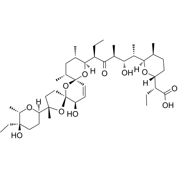
Salinomycin(Synonyms: 盐霉素; Procoxacin)

Salinomycin Chemical Structure

CAS No. : 53003-10-4

规格 价格 是否有货 数量
Free Sample (0.1-0.5 mg)   Apply now  
10 mM * 1 mL in DMSO ¥991 In-stock
5 mg ¥700 In-stock
10 mg ¥1200 In-stock
50 mg ¥3900 In-stock
100 mg   询价  
200 mg   询价  

* Please select Quantity before adding items.

Salinomycin 相关产品

相关化合物库:

  • Bioactive Compound Library Plus
  • Anti-Infection Compound Library
  • Apoptosis Compound Library
  • Stem Cell Signaling Compound Library
  • Wnt/Hedgehog/Notch Compound Library
  • Anti-Cancer Compound Library
  • Autophagy Compound Library
  • Anti-Aging Compound Library
  • Differentiation Inducing Compound Library
  • Antibacterial Compound Library
  • Cytoskeleton Compound Library
  • Antibiotics Library
  • Neuroprotective Compound Library
  • Anti-Breast Cancer Compound Library
  • Mitochondria-Targeted Compound Library
  • Transcription Factor Targeted Library
  • Anti-Liver Cancer Compound Library
  • Rare Diseases Drug Library
  • Anti-Colorectal Cancer Compound Library

生物活性

Salinomycin (Procoxacin), a polyether potassium ionophore antibiotic, selectively inhibits the growth of gram-positive bacteria. Salinomycin is a potent inhibitor of Wnt/β-catenin signaling, blocks Wnt-induced LRP6 phosphorylation. Salinomycin (Procoxacin) shows selective activity against human cancer stem cells[1][2][3].

体外研究
(In Vitro)

Salinomycin is a potent inhibitor of the Wnt signaling cascade. Incubation of the malignant lymphocytes with Salinomycin induces apoptosis within 48 h, with a mean IC50 of 230 nM. Salinomycin is also an antibiotic potassium ionophore, has been reported recently to act as a selective breast cancer stem cell inhibitor[1].
Salinomycin is a novel and an effective anticancer drug, inhibits SW620 cells and Cisp-resistant SW620 cells with IC50 of 1.54±0.23 μM and 0.32±0.05 μM, respectively. Salinomycin is found to have the ability to kill both cancer stem cells (CSCs) and therapy-resistant cancer cells. After continuous Salinomycin treatment for 48 h, the apoptotic cells are observed under the microscope and counted randomly at least 100 cells in one field. The number of apoptotic cells which are stained by Hoechst33342 is significantly increased in Cisp-resistant SW620 cells (20.20±3.72) than that of SW620 cells (9.40±2.07) per 100 cells (p<0.05). After treatment with Salinomycin for 48 h, flow cytometric analysis is used to detect the cell apoptosis both in SW620 cells and Cisp-resistant SW620 cells. The cell apoptotic rate in Cisp-resistant SW620 cells (37.82±3.63%) is significantly higher than that of SW620 cells (16.78±2.56%) (p<0.05)[2].

上海金畔生物科技有限公司 has not independently confirmed the accuracy of these methods. They are for reference only.

体内研究
(In Vivo)

After administration of 4 mg/kg Salinomycin (Sal), 8 mg/kg Salinomycin and 10 uL/g saline water for 6 weeks, the mice are sacrificed. The size of the liver tumors in the Salinomycin treatment groups diminishes compare with the control group. The mean diameter of the tumors decreases from 12.17 mm to 3.67 mm (p<0.05) and the mean volume (V=length×width2×0.5) of the tumors decreases from 819 mm3 to 25.25 mm3 (p<0.05). Next, the tumors are harvested, followed by HE staining, immunohistochemistry, and TUNEL assays, to assess the anti-tumor activity of Salinomycin. HE staining shows that the structure of the liver cancer tissue:nuclei of different sizes, hepatic cord structure is destroyed. Immunohistochemistry shows that PCNA expression is lower after Salinomycin treatment. HE staining and TUNEL assays indicates the Salinomycin-treated groups has higher apoptosis rates than control. Furthermore, immunohistochemistry shows an increased Bax/Bcl-2 ratio after Salinomycin treatment. The protein expression of β-catenin decreases in the Salinomycin treatment groups compared with control[4].
Salinomycin is a kind of monocarboxylic acid polyether type antibiotics, produced by the fermentation of Streptomyces albus, possesses a specific cyclic structure, and can form a complex compound with the pathogenic microorganisms and the extracellular cations of coccidian, especially K+, Na+, Rb+, to alter the intracellular and extracellular ion concentrations[5].

上海金畔生物科技有限公司 has not independently confirmed the accuracy of these methods. They are for reference only.

分子量

751.00

Formula

C42H70O11

CAS 号

53003-10-4

中文名称

盐霉素;沙利霉素

运输条件

Room temperature in continental US; may vary elsewhere.

储存方式
Powder -20°C 3 years
4°C 2 years
In solvent -80°C 6 months
-20°C 1 month
溶解性数据
In Vitro: 

DMSO : ≥ 36.7 mg/mL (48.87 mM)

* “≥” means soluble, but saturation unknown.

配制储备液
浓度 溶剂体积 质量 1 mg 5 mg 10 mg
1 mM 1.3316 mL 6.6578 mL 13.3156 mL
5 mM 0.2663 mL 1.3316 mL 2.6631 mL
10 mM 0.1332 mL 0.6658 mL 1.3316 mL

*

请根据产品在不同溶剂中的溶解度选择合适的溶剂配制储备液;一旦配成溶液,请分装保存,避免反复冻融造成的产品失效
储备液的保存方式和期限:-80°C, 6 months; -20°C, 1 month。-80°C 储存时,请在 6 个月内使用,-20°C 储存时,请在 1 个月内使用。

In Vivo:

请根据您的实验动物和给药方式选择适当的溶解方案。以下溶解方案都请先按照 In Vitro 方式配制澄清的储备液,再依次添加助溶剂:

——为保证实验结果的可靠性,澄清的储备液可以根据储存条件,适当保存;体内实验的工作液,建议您现用现配,当天使用; 以下溶剂前显示的百
分比是指该溶剂在您配制终溶液中的体积占比;如在配制过程中出现沉淀、析出现象,可以通过加热和/或超声的方式助溶

  • 1.

    请依序添加每种溶剂: 10% DMSO    40% PEG300    5% Tween-80    45% saline

    Solubility: ≥ 2.5 mg/mL (3.33 mM); Clear solution

    此方案可获得 ≥ 2.5 mg/mL (3.33 mM,饱和度未知) 的澄清溶液。

    以 1 mL 工作液为例,取 100 μL 25.0 mg/mL 的澄清 DMSO 储备液加到 400 μL PEG300 中,混合均匀;向上述体系中加入50 μL Tween-80,混合均匀;然后继续加入 450 μL生理盐水定容至 1 mL。

    将 0.9 g 氯化钠,完全溶解于 100 mL ddH₂O 中,得到澄清透明的生理盐水溶液

  • 2.

    请依序添加每种溶剂: 10% DMSO    90% corn oil

    Solubility: ≥ 2.5 mg/mL (3.33 mM); Clear solution

    此方案可获得 ≥ 2.5 mg/mL (3.33 mM,饱和度未知) 的澄清溶液,此方案不适用于实验周期在半个月以上的实验。

    以 1 mL 工作液为例,取 100 μL 25.0 mg/mL 的澄清 DMSO 储备液加到 900 μL玉米油中,混合均匀。

  • 3.

    请依序添加每种溶剂: 5% DMSO    40% PEG300    5% Tween-80    50% saline

    Solubility: 2.5 mg/mL (3.33 mM); Suspended solution; Need ultrasonic

*以上所有助溶剂都可在 上海金畔生物科技有限公司 网站选购。
参考文献
  • [1]. Lu D, et al. Salinomycin inhibits Wnt signaling and selectively induces apoptosis in chronic lymphocytic leukemia cells. Proc Natl Acad Sci U S A. 2011 Aug 9;108(32):13253-7.

    [2]. Zhou J, et al. Salinomycin induces apoptosis in cisplatin-resistant colorectal cancer cells by accumulation of reactiveoxygen species. Toxicol Lett. 2013 Oct 24;222(2):139-45.

    [3]. Klose J, et al. Salinomycin: Anti-tumor activity in a pre-clinical colorectal cancer model. PLoS One. 2019 Feb 14;14(2):e0211916.

    [4]. Wang F, et al. Salinomycin Inhibits Proliferation and Induces Apoptosis of Human Hepatocellular Carcinoma Cells In Vitro and In Vivo. PLoS One. 2012; 7(12): e50638.

    [5]. Qu H, et al. Effect of salinomycin on metastasis and invasion of bladder cancer cell line T24. Asian Pac J Trop Med. 2015 Jul;8(7):578-82.

    [6]. Naujokat C, et al. Salinomycin as a drug for targeting human cancer stem cells. J Biomed Biotechnol. 2012;2012:950658.

Cell Assay
[2]

For cisplatin or Salinomycin IC50 analysis in SW620 cells or Cisp-resistant SW620 cells, cells (1×10⁴/well) are cultured in 96-well plates and treated with different chemotherapeutics (cisplatin, Salinomycin) in different concentrations for 48 h. Then 20 μL of cell counting kit-8 (CCK-8) is added into each of the 96-wells. After 4 h incubation at 37°C, the optical density (OD) values are detected at 450 nm using the scan reader. Cell growth inhibiting rates are described as cell inhibiting curves and the IC50 parameters (inhibiting concentration of 50% cells) are evaluated by Xlfit 5.2 software. For cell proliferation analysis, SW620 cells or Cisp-resistant SW620 cells (5×10³/well) are also seeded in 96-well plates in serum-containing medium and treated with cisplatin (5 μM, according to the calculated IC50 values of cisplatin in SW620 cells) for 0, 12, 24, 48, 72 and 96 h. Then 20 μL cell counting kit-8 is added into each of the 96-wells. After 4-h incubation at 37°C, the coloring reactions are also quantified at 450 nm[2].

上海金畔生物科技有限公司 has not independently confirmed the accuracy of these methods. They are for reference only.

Animal Administration
[3][4]

Mice[3]
Nude mice (nu/nu; 4-6 weeks of age) are used. HepG2 cells are suspended in 100 mL 1:1 serum-free DMEM and Matrigel. Mice are anesthetized with ketamine/xylazine and after surgically opening the abdomen, HepG2 cells are inoculated into the liver parenchyma and mice are monitored every 3 days for 35 days. Finally, 18 nude mice are divided into three groups that are intraperitoneally injected daily for 6 weeks: two Salinomycin-treated groups (4 mg/kg Salinomycin group, 8 mg/kg Salinomycin group) and the control group (saline water group).
Rats[4]
A total of 10 male rats are used in the experiment. After a routine anesthesia, the abdomen is opened. After a resuspension of high glucose medium not containing serum DMEM, and matrigel, the bladder transitional cancer cell line T24 is inoculated in the parenchyma of bladder in rats, and then the abdomen is sutured. After operation, the rats are randomized into the experiment group and the control group with five in each group. After operation, the rats in the experiment group are immediately given intraperitoneal injection of Salinomycin with a dosage of 8 mg/kg, while the rats in the control group are given intraperitoneal injection of normal saline. A close observation is paid during the drug administration period. After 15 d, the rats are sacrificed by cervical dislocation, and the complete tumor tissues are stripped to observe the tumor growth and metastasis.

上海金畔生物科技有限公司 has not independently confirmed the accuracy of these methods. They are for reference only.

参考文献
  • [1]. Lu D, et al. Salinomycin inhibits Wnt signaling and selectively induces apoptosis in chronic lymphocytic leukemia cells. Proc Natl Acad Sci U S A. 2011 Aug 9;108(32):13253-7.

    [2]. Zhou J, et al. Salinomycin induces apoptosis in cisplatin-resistant colorectal cancer cells by accumulation of reactiveoxygen species. Toxicol Lett. 2013 Oct 24;222(2):139-45.

    [3]. Klose J, et al. Salinomycin: Anti-tumor activity in a pre-clinical colorectal cancer model. PLoS One. 2019 Feb 14;14(2):e0211916.

    [4]. Wang F, et al. Salinomycin Inhibits Proliferation and Induces Apoptosis of Human Hepatocellular Carcinoma Cells In Vitro and In Vivo. PLoS One. 2012; 7(12): e50638.

    [5]. Qu H, et al. Effect of salinomycin on metastasis and invasion of bladder cancer cell line T24. Asian Pac J Trop Med. 2015 Jul;8(7):578-82.

    [6]. Naujokat C, et al. Salinomycin as a drug for targeting human cancer stem cells. J Biomed Biotechnol. 2012;2012:950658.

所有产品仅用作科学研究或药证申报,我们不为任何个人用途提供产品和服务