上海金畔生物科技有限公司为生命科学和医药研发人员提供生物活性分子抑制剂、激动剂、特异性抑制剂、化合物库、重组蛋白,专注于信号通路和疾病研究领域。
CAL-130
CAL-130 是一种 PI3Kδ 和 PI3Kγ 抑制剂,IC50 分别为 1.3 和 6.1 nM。

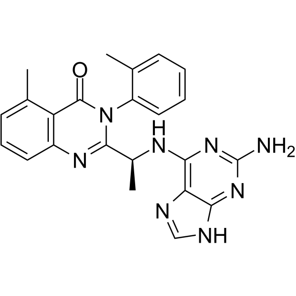
CAL-130 Chemical Structure
CAS No. : 1431697-74-3
| 规格 | 是否有货 | ||
|---|---|---|---|
| 100 mg | 询价 | ||
| 250 mg | 询价 | ||
| 500 mg | 询价 |
* Please select Quantity before adding items.
CAL-130 的其他形式现货产品:
| 生物活性 |
CAL-130 is a PI3Kδ and PI3Kγ inhibitor with IC50s of 1.3 and 6.1 nM, respectively. |
||||
|---|---|---|---|---|---|
| IC50 & Target[1] |
|
||||
| 体外研究 (In Vitro) |
CAL-130 preferentially inhibits the function of both p110γ and p110δ catalytic domains. IC50 values of CAL-130 are 1.3 and 6.1 nM for p110δ and p110γ, respectively, as compared to 115 and 56 nM for p110α and p110β. CAL-130 does not inhibit additional intracellular signaling pathways (i.e., p38 MAPK or insulin receptor tyrosine kinase) that are critical for general cell function and survival[1]. 上海金畔生物科技有限公司 has not independently confirmed the accuracy of these methods. They are for reference only. |
||||
| 体内研究 (In Vivo) |
The clinical significance of interfering with the combined activities of PI3Kγ and PI3Kδ is determined by administering CAL-130 to Lck/Ptenfl/fl mice with established T cell acute lymphoblastic leukemia (T-ALL). Candidate animals for survival studies are ill appearing, have a white blood cell (WBC) count above 45,000 μL-1, evidence of blasts on peripheral smear, and a majority of circulation cells (>75%) staining double positive for Thy1.2 and Ki-67. Mice receive an oral dose (10 mg/kg) of CAL-130 every 8 hr for a period of 7 days and are then followed until moribund. Despite the limited duration of therapy, CAL-130 is highly effective in extending the median survival for treated animals to 45 days as compared 7.5 days for the control group[1]. 上海金畔生物科技有限公司 has not independently confirmed the accuracy of these methods. They are for reference only. |
||||
| 分子量 |
426.47 |
||||
| Formula |
C23H22N8O |
||||
| CAS 号 |
1431697-74-3 |
||||
| 运输条件 |
Room temperature in continental US; may vary elsewhere. |
||||
| 储存方式 |
Please store the product under the recommended conditions in the Certificate of Analysis. |
||||
| 参考文献 |
|
| Kinase Assay [1] |
IC50 values for CAL-130 inhibition of PI3K isoforms are determined in ex vivo PI3 kinase assays using recombinant PI3K. A ten-point kinase inhibitory profile is determined with ATP at a concentration consistent with the KM for each enzyme[1]. 上海金畔生物科技有限公司 has not independently confirmed the accuracy of these methods. They are for reference only. |
|---|---|
| Cell Assay [1] |
Cell proliferation of CCRF-CEM cells or shRNA-transfected CCRF-CEM cells, in presence or absence of CAL-130 (1, 2.5 and 5 μM), is followed by cell counting of samples in triplicate using a hemocytometer and trypan blue. For apoptosis determinations of untransfected or shRNA-transfected CCRF-CEMs, cells are stained with APC-conjugated Annexin-V in Annexin Binding Buffer and analyzed by flow cytometry. For primary T-ALL samples, cell viability is assessed using the BD Cell Viability kit coupled with the use of fluorescentcounting beads. For this, cells are plated with MS5-DL1 stroma cells, and after 72 hr following CAL-130 treatment, cells are harvested and stained with an APC-conjugated antihuman CD45 followed by a staining with the aforementioned kit[1]. 上海金畔生物科技有限公司 has not independently confirmed the accuracy of these methods. They are for reference only. |
| Animal Administration [1] |
Mice[1] 上海金畔生物科技有限公司 has not independently confirmed the accuracy of these methods. They are for reference only. |
| 参考文献 |
|
所有产品仅用作科学研究或药证申报,我们不为任何个人用途提供产品和服务
