上海金畔生物科技有限公司为生命科学和医药研发人员提供生物活性分子抑制剂、激动剂、特异性抑制剂、化合物库、重组蛋白,专注于信号通路和疾病研究领域。
Ciliobrevin A (Synonyms: HPI-4) 纯度: 98.72%
Ciliobrevin A (HPI-4) 是一种 Hedgehog (Hh) 信号通路抑制剂,平均抑制浓度 (IC50) 值小于 10 μM。

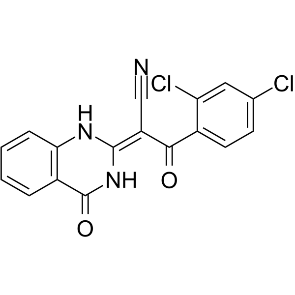
Ciliobrevin A Chemical Structure
CAS No. : 302803-72-1
| 规格 | 价格 | 是否有货 | 数量 |
|---|---|---|---|
| 10 mM * 1 mL in DMSO | ¥638 | In-stock | |
| 5 mg | ¥580 | In-stock | |
| 10 mg | ¥770 | In-stock | |
| 25 mg | ¥1700 | In-stock | |
| 50 mg | ¥3100 | In-stock | |
| 100 mg | ¥4950 | In-stock | |
| 200 mg | 询价 | ||
| 500 mg | 询价 |
* Please select Quantity before adding items.
Ciliobrevin A 相关产品
•相关化合物库:
- Bioactive Compound Library Plus
- Stem Cell Signaling Compound Library
- Wnt/Hedgehog/Notch Compound Library
- Anti-Cancer Compound Library
- Anti-Aging Compound Library
- Differentiation Inducing Compound Library
- Anti-Breast Cancer Compound Library
- Anti-Pancreatic Cancer Compound Library
- Anti-Blood Cancer Compound Library
- Targeted Diversity Library
- Anti-Liver Cancer Compound Library
- Anti-Colorectal Cancer Compound Library
| 生物活性 |
Ciliobrevin A (HPI-4) is a hedgehog (Hh) signaling pathway inhibitor with median inhibitory concentration (IC50) less than 10 μM[1]. |
IC50 & Target |
IC50: <10 μm (hedgehog)[1] |
||||||||||||||
|---|---|---|---|---|---|---|---|---|---|---|---|---|---|---|---|---|---|
| 体外研究 (In Vitro) |
Ciliobrevin A (HPI-4) also prevents an increase in the FLAG-Gli2 full-length/repressor ratio upon Shh stimulation, but HPI-2 and HPI-3 have no significant effect. Ciliobrevin A decreases FLAG-Gli1 stability in these cells, revealing another mechanism by which this small molecule can inhibit Hh target gene expression, while neither HPI-2 or HPI-3 has any significant effect on FLAG-Gli1 levels. Ciliobrevin A increases ciliary levels of FLAG-Gli2 in a manner disproportionate to their effects on total FLAG-Gli2 levels. In addition, Shh-EGFPFLAG-Gli2 cells cultured with Ciliobrevin A have truncated primary cilia, and this cellular organelle is absent in a significant fraction of Ciliobrevin A-treated cells. Ciliobrevin A also perturbs primary cilia formation in the Shh-LIGHT2FLAG-Gli1 cells and promotes accumulation of FLAG-Gli1 at the distal tip of this organelle. Ciliobrevin A significantly inhibits the proliferation of these neuronal progenitors, as measured by histone H3 phosphorylation (pH3) levels, and reduces cellular levels of cyclin D1 protein and Gli1, Gli2, and N-Myc transcripts in the CGNPs. Ciliobrevin A can block the proliferation of SmoM2-expressing CGNPs and should be equally potent against CGNPs lacking Su(fu) function, whereas the Smo inhibitor Cyclopamine is ineffective against either oncogenic lesion[1]. 上海金畔生物科技有限公司 has not independently confirmed the accuracy of these methods. They are for reference only. |
||||||||||||||||
| 分子量 |
358.18 |
||||||||||||||||
| Formula |
C17H9Cl2N3O2 |
||||||||||||||||
| CAS 号 |
302803-72-1 |
||||||||||||||||
| 运输条件 |
Room temperature in continental US; may vary elsewhere. |
||||||||||||||||
| 储存方式 |
|
||||||||||||||||
| 溶解性数据 |
In Vitro:
DMSO : 100 mg/mL (279.19 mM; Need ultrasonic) 配制储备液
*
请根据产品在不同溶剂中的溶解度选择合适的溶剂配制储备液;一旦配成溶液,请分装保存,避免反复冻融造成的产品失效。 In Vivo:
请根据您的实验动物和给药方式选择适当的溶解方案。以下溶解方案都请先按照 In Vitro 方式配制澄清的储备液,再依次添加助溶剂: ——为保证实验结果的可靠性,澄清的储备液可以根据储存条件,适当保存;体内实验的工作液,建议您现用现配,当天使用; 以下溶剂前显示的百
|
||||||||||||||||
| 参考文献 |
|
| Kinase Assay [1] |
Smo-binding assays are conducted with BODIPY-cyclopamine and Smo-overexpressing HEK 293T cells, using a CMVpromoter-based SV40 origin-containing expression construct for Smo-Myc3 (murine Smo containing three consecutive Myc epitopes at the C terminus). HEK 293T cells are seeded into eight-well chambered coverslips (80,000 cells/well) and cultured in DMEM containing 10% FBS, 100 U/mL penicillin, and 0.1 mg/mL streptomycin. The cells are cultured until they reached 55 to 65% confluency (14-18 h), after which they are transfected with the Smo-Myc3 expression construct and Transit-LT1. Twenty-four hours after transfection, the cells are washed with PBS and cultured in DMEM containing 0.5% FBS, 5 nM BODIPY-cyclopamine, and various concentrations of either cyclopamine or individual HPIs. After 30 min, 10 μM Hoescht 33342 is added to each well, and the HPIs are incubated with the cells for an additional 30 min. The cells are then washed two times with PBS buffer, once with phenol red-free DMEM containing 0.5% FBS, and immediately imaged using a DMI6000B compound microscope. Images are background-substracted using ImageJ software with a rolling ball size of 75 pixels, and BODIPY-cyclopamine intensity is then determined using Metamorph software. Circular regions with a diameter of 300 pixels are placed over regions containing uniformly confluent cells, and the pixel intensities of approximately 20 regions from four independent images is used to determine the average BODIPY-cyclopamine levels for each experimental condition[1]. 上海金畔生物科技有限公司 has not independently confirmed the accuracy of these methods. They are for reference only. |
|---|---|
| Cell Assay [1] |
NIH 3T3 cells are seeded into 24-well plates (40,000 cell/well) containing polyD-lysine-coated 12-mm glass coverslips and cultured in DMEM containing 10% CS, 100 U/mL penicillin, and 0.1 mg/mL streptomycin until they reached 85-90% confluency. The medium is changed to DMEM containing 0.5% CS, 100 U/mL penicillin, and 0.1 mg/mL streptomycin and the cells are cultured for another 12 h. The cells are then treated with either DMSO, 3 μM cyclopamine,15 μM HPI-1, 20 μM HPI-2, 30 μM HPI-3, or 30 μM Ciliobrevin A. Shh-N-conditioned medium is added to appropriate wells at a final concentration of 5%. After 12 h, the cells are fixed in 4% paraformaldehyde for 10 min at room temperature, washed three times with PBS, permeabilized for 1 min with PBS containing 0.1% Triton X-100, washed again three times with PBS, and then blocked with PBS containing 1% normal goat serum for 3 h. The coverslips are then treated with mouse anti-N-acetylated-α-tubulin (1:1,000 in blocking buffer) and rabbit anti-Smo antibody (6) (1:2,000 dilution in blocking buffer) for 2 h at room temperature and washed 3×5 min with PBS. The coverslips are incubated next with Alexa Fluor 594-conjugated goat anti-mouse IgG and Alexa Fluor 488-conjugated donkey anti-rabbit IgG antibodies (1:1,000 dilutions in blocking buffer) for 1 h at room temperature. After washes with PBS and a 5-min incubation with 4,6-diamidino-2-phenylindole (DAPI), the samples are mounted using Prolong Gold and imaged with an inverted Leica DMIRE2 laser scaning confocal microscope[1]. 上海金畔生物科技有限公司 has not independently confirmed the accuracy of these methods. They are for reference only. |
| 参考文献 |
|
所有产品仅用作科学研究或药证申报,我们不为任何个人用途提供产品和服务
