上海金畔生物科技有限公司为生命科学和医药研发人员提供生物活性分子抑制剂、激动剂、特异性抑制剂、化合物库、重组蛋白,专注于信号通路和疾病研究领域。
FRAX1036 纯度: 98.06%
FRAX1036 是一种 PAK 抑制剂,对 PAK1,PAK2 和 PAK4 的 Ki 值分别为 23.3 nM,72.4 nM 和 2.4 μM。

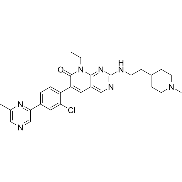
FRAX1036 Chemical Structure
CAS No. : 1432908-05-8
| 规格 | 价格 | 是否有货 | 数量 |
|---|---|---|---|
| 10 mM * 1 mL in DMSO | ¥1596 | In-stock | |
| 1 mg | ¥700 | In-stock | |
| 5 mg | ¥1400 | In-stock | |
| 10 mg | ¥2200 | In-stock | |
| 50 mg | ¥8800 | In-stock | |
| 100 mg | ¥14000 | In-stock | |
| 200 mg | 询价 | ||
| 500 mg | 询价 |
* Please select Quantity before adding items.
FRAX1036 相关产品
•相关化合物库:
- Bioactive Compound Library Plus
- Cell Cycle/DNA Damage Compound Library
- Kinase Inhibitor Library
- Anti-Cancer Compound Library
- Anti-Aging Compound Library
- Cytoskeleton Compound Library
- Anti-Lung Cancer Compound Library
| 生物活性 |
FRAX1036 is a PAK inhibitor with Kis of 23.3 nM, 72.4 nM, and 2.4 μM for PAK1, PAK2 and PAK4, respectively. |
||||||||||||||||
|---|---|---|---|---|---|---|---|---|---|---|---|---|---|---|---|---|---|
| IC50 & Target[1] |
|
||||||||||||||||
| 体外研究 (In Vitro) |
FRAX1036 is a PAK inhibitor with Kis of 23.3 nM, 72.4 nM, and 2.4 μM for PAK1, PAK2 and PAK4, respectively. FRAX1036 (2.5 μM) incombination with docetaxel alters stathmin phosphorylation, induces the apoptotic marker cleaved PARP and increases kinetics of apoptosis in MDA-MB-175 and HCC2911 cells; also alters microtubule organization, mitosis and cell fate in U2OS cells. Moreover, FRAX1036 shows significantly effective inhibition on U2OS cells[1]. FRAX1036 (10 μM) affects the proliferation of non-small cell lung cancer (NSCLC) cells when added to KRAS prenylation inhibitors[2]. 上海金畔生物科技有限公司 has not independently confirmed the accuracy of these methods. They are for reference only. |
||||||||||||||||
| 分子量 |
518.05 |
||||||||||||||||
| Formula |
C28H32ClN7O |
||||||||||||||||
| CAS 号 |
1432908-05-8 |
||||||||||||||||
| 运输条件 |
Room temperature in continental US; may vary elsewhere. |
||||||||||||||||
| 储存方式 |
|
||||||||||||||||
| 溶解性数据 |
In Vitro:
DMSO : 5.3 mg/mL (10.23 mM; Need warming) 配制储备液
*
请根据产品在不同溶剂中的溶解度选择合适的溶剂配制储备液;一旦配成溶液,请分装保存,避免反复冻融造成的产品失效。 |
||||||||||||||||
| 参考文献 |
|
| Kinase Assay [1] |
The activity/inhibition of human recombinant PAK1 (kinase domain), PAK2 (full length) or PAK4 (kinase domain) is estimated by measuring the phosphorylation of a FRET peptide substrate (Ser/Thr19) labeled with Coumarin and Fluorescein using Z’-LYTETM assay. The 10 μL assay mixtures containe 50 mM HEPES (pH 7.5), 0.01% Brij-35, 10 mM MgCl2, 1 mM EGTA, 2 μM FRET peptide substrate, and PAK enzyme (20 pM PAK1; 50 pM PAK2; 90 pM PAK4). Incubations are carried out at 22°C in black polypropylene 384-well plates. Prior to the assay, enzyme, FRET peptide substrate and serially diluted test compounds (FRAX1036, etc.) are preincubated together in assay buffer (7.5 μL) for 10 minutes, and the assay is initiated by the addition of 2.5 μL assay buffer containing 4× ATP (160 μM PAK1; 480 μM PAK2; 16 μM PAK4). Following the 60-minute incubation, the assay mixtures are quenched by the addition of 5 μL of Z’-LYTETM development reagent, and 1 hour later the emissions of Coumarin (445 nm) and Fluorescein (520 nm) are determined after excitation at 400 nm. An emission ratio (445 nm/520 nm) is determined to quantify the degree of substrate phosphorylation[1]. 上海金畔生物科技有限公司 has not independently confirmed the accuracy of these methods. They are for reference only. |
|---|---|
| Cell Assay [1] |
For caspase 3/7 activation apoptosis assays, cells are plated at 10,000 cells/well in 96-well plates for 24 hours prior to treating with DMSO, FRAX1036, and/or docetaxel. Caspase 3/7 reagent is added at a 1:1000 dilution. Cells are imaged at 10× magnification in an IncuCyte Zoom Live-content imaging system at 37°C, 5% CO2. Images are acquired every 2 hours or 4 hours for 36 to 72 hours, two images/well. Data is analyzed using IncuCyte analysis software to detect and quantify green (apoptotic) cells/image. Each condition is performed in triplicate. Averages with SEM at each time point are plotted in Excel. A t-test is performed for the final time point comparing the combination of FRAX1036 and docetaxel with each single agent in Prism. The apoptotic index is calculated from the apoptosis assays by dividing the final apoptotic cell count by the total cell count. Averages with SEM are plotted in Excel, and a t-test is performed comparing the combination of FRAX1036 and docetaxel with each single agent in Prism[1]. 上海金畔生物科技有限公司 has not independently confirmed the accuracy of these methods. They are for reference only. |
| 参考文献 |
|
所有产品仅用作科学研究或药证申报,我们不为任何个人用途提供产品和服务
