CNDAC

上海金畔生物科技有限公司为生命科学和医药研发人员提供生物活性分子抑制剂、激动剂、特异性抑制剂、化合物库、重组蛋白,专注于信号通路和疾病研究领域。

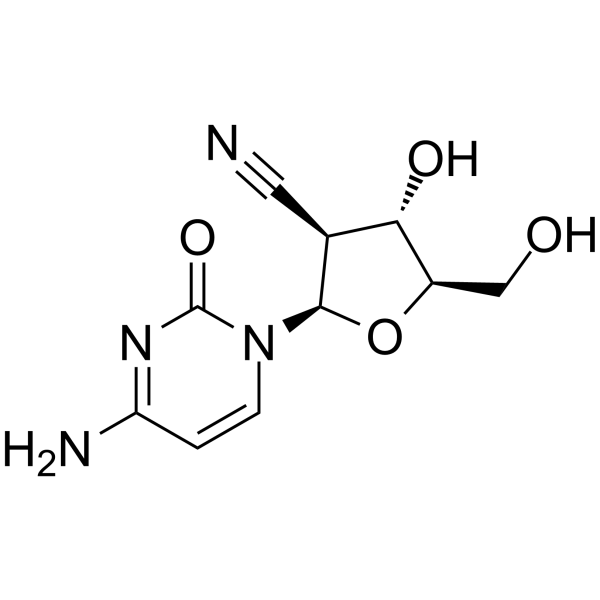
CNDAC 

CNDAC 是 sapacitabine 的有效代谢物,为一种核苷类似物。

CNDAC

CNDAC Chemical Structure

CAS No. : 135598-68-4

规格 是否有货
100 mg   询价  
250 mg   询价  
500 mg   询价  

* Please select Quantity before adding items.

CNDAC 的其他形式现货产品:

Sapacitabine CNDAC hydrochloride

生物活性

CNDAC is a major metabolite of oral drug sapacitabine, and a nucleoside analog.

体外研究
(In Vitro)

CNDAC-induced SSBs can be repaired by the transcription-coupled nucleotide excision repair pathway, whereas lethal DSBs are mainly repaired through homologous recombination. Deficiency in two Rad51 paralogs, Rad51D and XRCC3, greatly sensitize cells to CNDAC. The Rad51D-null cell line is approximately 50-fold more sensitive to CNDAC (IC50=0.006 µM) compared to 51D1.3, the Rad51D-repleted line (IC50=0.32 µM)[1]. CNDAC shows inhibitory activity against HL-60 and THP-1 cells with IC50s of 1.58 µM and 0.84 µM. CNDAC (10 μM) results in a significant drop in cell survival compared to the untreated on days 4, 7, and 14. CNDAC is more effective at reducing viability and inducing apoptosis than ara-C at equivalent concentrations in the THP-1 cell line, which is defined as displaying resistance to ara-C[2]. CNDAC induces DSBs, which are products of replication, rather than a consequence of induction of apoptosis. CNDAC causes DNA damage, and DNA-PK and ATR are dispensable for cell survival. CNDAC exhibits potent activity against human fibroblasts deficient in ATM or transfected with an empty vector, approximately 30-fold more than cells repleted with full-length ATM cDNA, with IC50s of 0.01 μM and 0.3 μM, respectively. CNDAC-induced DNA damage is repaired through the homologous recombination pathway[3].

Shanghai Jinpan Biotech Co Ltd has not independently confirmed the accuracy of these methods. They are for reference only.

分子量

252.23

Formula

C10H12N4O4

CAS 号

135598-68-4

运输条件

Room temperature in continental US; may vary elsewhere.

储存方式

Please store the product under the recommended conditions in the Certificate of Analysis.

参考文献
  • [1]. Liu XJ, et al. Sapacitabine, the prodrug of CNDAC, is a nucleoside analog with a unique action mechanism of inducing DNA strand breaks. hin J Cancer. 2012 Aug;31(8):373-80.

    [2]. Jagan S, et al. Bone Marrow and Peripheral Blood AML Cells Are Highly Sensitive to CNDAC, the Active Form of Sapacitabine. Adv Hematol. 2012;2012:727683.

    [3]. Liu X, et al. Homologous recombination as a resistance mechanism to replication-induced double-strand breaks caused by the antileukemia agent CNDAC. Blood. 2010 Sep 9;116(10):1737-46.

Cell Assay
[2]

1×10⁶ primary BM and PB cells are treated with 1 μM (low), 10 μM (medium), and 100 μM (high) of ara-C or CNDAC or 0.005 μM (low), 0.05 μM (medium) and 0.5 μM (high) mitoxantrone in 24 well plates at 37°C, 5% CO2, and 100% humidity for 4 days. Appropriate untreated controls are included. Postdrug treatment, both PB and BM non-adherent cells are washed to remove compound, replated on M2-10B4 stromal layers, and reincubated at 37°C, 5% CO2, 100% humidity. Cells are analyzed immediately posttreatment and following 3, 7, and 31 days postdrug removal.

Shanghai Jinpan Biotech Co Ltd has not independently confirmed the accuracy of these methods. They are for reference only.

参考文献
  • [1]. Liu XJ, et al. Sapacitabine, the prodrug of CNDAC, is a nucleoside analog with a unique action mechanism of inducing DNA strand breaks. hin J Cancer. 2012 Aug;31(8):373-80.

    [2]. Jagan S, et al. Bone Marrow and Peripheral Blood AML Cells Are Highly Sensitive to CNDAC, the Active Form of Sapacitabine. Adv Hematol. 2012;2012:727683.

    [3]. Liu X, et al. Homologous recombination as a resistance mechanism to replication-induced double-strand breaks caused by the antileukemia agent CNDAC. Blood. 2010 Sep 9;116(10):1737-46.

所有产品仅用作科学研究或药证申报,我们不为任何个人用途提供产品和服务
Inolitazone(Synonyms: Efatutazone; CS-7017; RS5444)

上海金畔生物科技有限公司为生命科学和医药研发人员提供生物活性分子抑制剂、激动剂、特异性抑制剂、化合物库、重组蛋白,专注于信号通路和疾病研究领域。

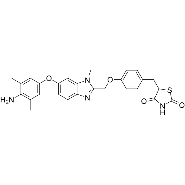
Inolitazone (Synonyms: Efatutazone; CS-7017; RS5444)

Inolitazone(RS5444; CS-7017)是新型PPARγ高亲和性激动剂,EC50为rosiglitazone的五十分之一,对于不表达PPARγ的RIE细胞无抑制性。

Inolitazone(Synonyms: Efatutazone;  CS-7017;  RS5444)

Inolitazone Chemical Structure

CAS No. : 223132-37-4

规格 是否有货
100 mg   询价  
250 mg   询价  
500 mg   询价  

* Please select Quantity before adding items.

Inolitazone 的其他形式现货产品:

Inolitazone dihydrochloride

生物活性

Inolitazone a novel high-affinity PPARγ agonist that is dependent upon PPARγ for its biological activity with IC50 of 0.8 nM for growth inhibition.

IC50 & Target

PPARγ


体外研究
(In Vitro)

Inolitazone (RS5444) upregulates the cell cycle kinase inhibitor, p21 WAF1/CIP1. Silencing p21 WAF1/CIP1 rendered cells insensitive to Inolitazone. A 10 nM dose of Inolitazone activates PPARγ:RXRα-dependent transcription as demonstrated in a transient transfection assay utilizing a PPRE response element fused to a luciferase reporter gene (PPRE3-tk-luc). DRO cells are treated in culture with Inolitazone, Rosiglitazone, or Troglitazone at the indicated concentrations. DRO cells are transiently transfected with PPRE3-tk-luc to examine effective concentrations at which EC50 occurs. The EC50s are 1 nM (Inolitazone), 65 nM (Rosiglitazone) and 631 nM (Troglitazone). Similarly, the calculated inhibitory concentration at IC50 is 0.8 nM for Inolitazone, 75 nM for Rosiglitazone, and 1412 nM for Troglitazone. Inolitazone specifically activates PPARγ, but not PPARα or PPARδ. Exposure of 10 nM Inolitazone following transient transfection with the appropriate PPAR isoform (γ, α, or δ) and PPAR response element linked to a luciferase reporter in RIE rat small intestinal cell line, which does not express PPARs, yields increased luciferase activity only in the presence of PPARγ and PPRE3-tk-luc[1]. DRO cells are growth inhibited by 10 nM Inolitazone (RS5444) through a PPARγ-dependent mechanism[2].

Shanghai Jinpan Biotech Co Ltd has not independently confirmed the accuracy of these methods. They are for reference only.

体内研究
(In Vivo)

Inolitazone (RS5444) plus Paclitaxel demonstrate additive antiproliferative activity in cell culture and minimal ATC tumor growth. When Inolitazone is administered in the diet to athymic nude mice prior to DRO tumor cell implantation, tumor growth is inhibited in a dose responsive fashion. At the highest dose, 0.025% Inolitazone inhibits growth on day 32 by 94.4% as compared to that of control. In this treatment group, five of 10 animals do not develop demonstrable tumors. In the 0.0025% treatment group, tumor growth is inhibited by 62.3% compared to that of control on day 32 while the 0.00025% dose demonstrated no growth inhibitory activity as compared to control. Tumors is nest allowed to establish in the mouse and began 0.025% Inolitazone treatment of mice 1 week after DRO or ARO tumor cell implantation. Inolitazone treated animals demonstrate tumor growth inhibition of 68.9% in DRO tumors and 48.3% in ARO tumors as compared to that of their respective controls on day 35[1].

Shanghai Jinpan Biotech Co Ltd has not independently confirmed the accuracy of these methods. They are for reference only.

Clinical Trial

分子量

502.58

Formula

C27H26N4O4S

CAS 号

223132-37-4

中文名称

伊诺他酮二盐酸盐

运输条件

Room temperature in continental US; may vary elsewhere.

储存方式

Please store the product under the recommended conditions in the Certificate of Analysis.

参考文献
  • [1]. Copland JA, et al. Novel high-affinity PPARgamma agonist alone and in combination with paclitaxel inhibits human anaplastic thyroid carcinoma tumor growth via p21WAF1/CIP1. Oncogene. 2006 Apr 13;25(16):2304-17.

    [2]. Marlow LA, et al. Reactivation of suppressed RhoB is a critical step for the inhibition of anaplastic thyroid cancer growth. Cancer Res. 2009 Feb 15;69(4):1536-44.

Cell Assay
[1]

DRO90-1 (DRO) and ARO81 (ARO) cells are plated in 12-well culture plates in triplicate for each condition at an initial concentration of 2×10⁴ cells/well. After overnight incubation, cells are treated with either Inolitazone, Rosiglitazone, Troglitazone, GW9662, or Paclitaxel diluted in DMSO at concentrations indicated in figure legends. All cells receive identical volumes of DMSO and are exposed to each drug for 6 days with medium and drug changed every 48 h. After 6 days, cells are washed with PBS, trypsinized and counted by Beckman Coulter Counter[1].

Shanghai Jinpan Biotech Co Ltd has not independently confirmed the accuracy of these methods. They are for reference only.

Animal Administration
[1]

Mice[1]
Suspensions of 1×10⁶/0.1 mL DRO or ARO cells in RPMI medium are injected subcutaneously in one flank of 3-4 week athymic female nude mice. Mice are changed to specialized diets either 1 week prior or 1 week after tumor implantation and randomly assigned to experimental or control groups with 10 mice per group. Diets consisted either placebo, 0.00025%, 0.0025%, or 0.025% Inolitazone formulated into the diet. Mice weighed between 20-25 g and consume on average 4 g of food per day. For combinatorial studies either placebo, 10 mg/kg or 15 mg/kg paclitaxel is injected i.p. twice weekly. Tumors are measured every 3-4 days for 35 days with calipers and tumor volumes are calculated by the formula: 0.5236(a×b×c), where a is the shortest diameter, b is the diameter perpendicular to a and c is the diameter height.

Shanghai Jinpan Biotech Co Ltd has not independently confirmed the accuracy of these methods. They are for reference only.

参考文献
  • [1]. Copland JA, et al. Novel high-affinity PPARgamma agonist alone and in combination with paclitaxel inhibits human anaplastic thyroid carcinoma tumor growth via p21WAF1/CIP1. Oncogene. 2006 Apr 13;25(16):2304-17.

    [2]. Marlow LA, et al. Reactivation of suppressed RhoB is a critical step for the inhibition of anaplastic thyroid cancer growth. Cancer Res. 2009 Feb 15;69(4):1536-44.

所有产品仅用作科学研究或药证申报,我们不为任何个人用途提供产品和服务

CNDAC

上海金畔生物科技有限公司为生命科学和医药研发人员提供生物活性分子抑制剂、激动剂、特异性抑制剂、化合物库、重组蛋白,专注于信号通路和疾病研究领域。

CNDAC 

CNDAC 是 sapacitabine 的有效代谢物,为一种核苷类似物。

CNDAC

CNDAC Chemical Structure

CAS No. : 135598-68-4

规格 是否有货
100 mg   询价  
250 mg   询价  
500 mg   询价  

* Please select Quantity before adding items.

CNDAC 的其他形式现货产品:

Sapacitabine CNDAC hydrochloride

生物活性

CNDAC is a major metabolite of oral drug sapacitabine, and a nucleoside analog.

体外研究
(In Vitro)

CNDAC-induced SSBs can be repaired by the transcription-coupled nucleotide excision repair pathway, whereas lethal DSBs are mainly repaired through homologous recombination. Deficiency in two Rad51 paralogs, Rad51D and XRCC3, greatly sensitize cells to CNDAC. The Rad51D-null cell line is approximately 50-fold more sensitive to CNDAC (IC50=0.006 µM) compared to 51D1.3, the Rad51D-repleted line (IC50=0.32 µM)[1]. CNDAC shows inhibitory activity against HL-60 and THP-1 cells with IC50s of 1.58 µM and 0.84 µM. CNDAC (10 μM) results in a significant drop in cell survival compared to the untreated on days 4, 7, and 14. CNDAC is more effective at reducing viability and inducing apoptosis than ara-C at equivalent concentrations in the THP-1 cell line, which is defined as displaying resistance to ara-C[2]. CNDAC induces DSBs, which are products of replication, rather than a consequence of induction of apoptosis. CNDAC causes DNA damage, and DNA-PK and ATR are dispensable for cell survival. CNDAC exhibits potent activity against human fibroblasts deficient in ATM or transfected with an empty vector, approximately 30-fold more than cells repleted with full-length ATM cDNA, with IC50s of 0.01 μM and 0.3 μM, respectively. CNDAC-induced DNA damage is repaired through the homologous recombination pathway[3].

上海金畔生物科技有限公司 has not independently confirmed the accuracy of these methods. They are for reference only.

分子量

252.23

Formula

C10H12N4O4

CAS 号

135598-68-4

运输条件

Room temperature in continental US; may vary elsewhere.

储存方式

Please store the product under the recommended conditions in the Certificate of Analysis.

参考文献
  • [1]. Liu XJ, et al. Sapacitabine, the prodrug of CNDAC, is a nucleoside analog with a unique action mechanism of inducing DNA strand breaks. hin J Cancer. 2012 Aug;31(8):373-80.

    [2]. Jagan S, et al. Bone Marrow and Peripheral Blood AML Cells Are Highly Sensitive to CNDAC, the Active Form of Sapacitabine. Adv Hematol. 2012;2012:727683.

    [3]. Liu X, et al. Homologous recombination as a resistance mechanism to replication-induced double-strand breaks caused by the antileukemia agent CNDAC. Blood. 2010 Sep 9;116(10):1737-46.

Cell Assay
[2]

1×10⁶ primary BM and PB cells are treated with 1 μM (low), 10 μM (medium), and 100 μM (high) of ara-C or CNDAC or 0.005 μM (low), 0.05 μM (medium) and 0.5 μM (high) mitoxantrone in 24 well plates at 37°C, 5% CO2, and 100% humidity for 4 days. Appropriate untreated controls are included. Postdrug treatment, both PB and BM non-adherent cells are washed to remove compound, replated on M2-10B4 stromal layers, and reincubated at 37°C, 5% CO2, 100% humidity. Cells are analyzed immediately posttreatment and following 3, 7, and 31 days postdrug removal.

上海金畔生物科技有限公司 has not independently confirmed the accuracy of these methods. They are for reference only.

参考文献
  • [1]. Liu XJ, et al. Sapacitabine, the prodrug of CNDAC, is a nucleoside analog with a unique action mechanism of inducing DNA strand breaks. hin J Cancer. 2012 Aug;31(8):373-80.

    [2]. Jagan S, et al. Bone Marrow and Peripheral Blood AML Cells Are Highly Sensitive to CNDAC, the Active Form of Sapacitabine. Adv Hematol. 2012;2012:727683.

    [3]. Liu X, et al. Homologous recombination as a resistance mechanism to replication-induced double-strand breaks caused by the antileukemia agent CNDAC. Blood. 2010 Sep 9;116(10):1737-46.

所有产品仅用作科学研究或药证申报,我们不为任何个人用途提供产品和服务

郑州长城科工贸GRS系列防爆调速双层玻璃反应釜GRS-20EX

郑州长城科工贸GRS系列防爆调速双层玻璃反应釜GRS-20EX

  • 品牌 长城科工贸|Greatwall
  • 型号 GRS-20EX
  • 商品详情

    专利号:201630293823.8

    用途特点:

    ●框架为不锈钢材质,防腐蚀。

    ●配置316材质接料托盘,可回收物料,防止污染环境。

    ●高硼硅玻璃材质,具有优良的物理、化学性能。

    ●可在高温(200℃)至低温(-80℃)的范围内使用。

    ●可在常压及负压条件下工作,真空度可达0.095MPa。

    ●釜内可承受压力范围:-0.1MPa~0MPa。

    ●特氟隆+FV橡胶材质的旋塞或放料阀;外加四氟乙烯保护层的“O”型圈。

    型号

    GRS-20

    GRS-20EX

    功率(减速比:3)(W)

    90

    180

    搅拌转速(rpm)

    50~500

    最大扭矩(N*m)

    0.6

    物料容积(L)

    20

    夹套容积(L)

    6

    电源

    220-240V~,50/60Hz

    放料阀离地高(mm)

    550

    整机高度(mm)

    2135


    型号

    GRS-50

    GRS-50EX

    功率(减速比:3)(W)

    140

    180

    搅拌转速(rpm)

    50~500

    最大扭矩(N*m)

    0.95

    物料容积(L)

    50

    夹套容积(L)

    16

    电源

    220-240V~,50/60Hz

    放料阀离地高(mm)

    550

    整机高度(mm)

    2650

  • CNDAC

    上海金畔生物科技有限公司为生命科学和医药研发人员提供生物活性分子抑制剂、激动剂、特异性抑制剂、化合物库、重组蛋白,专注于信号通路和疾病研究领域。

    CNDAC 

    CNDAC 是 sapacitabine 的有效代谢物,为一种核苷类似物。

    CNDAC

    CNDAC Chemical Structure

    CAS No. : 135598-68-4

    规格 是否有货
    100 mg   询价  
    250 mg   询价  
    500 mg   询价  

    * Please select Quantity before adding items.

    CNDAC 的其他形式现货产品:

    Sapacitabine CNDAC hydrochloride

    生物活性

    CNDAC is a major metabolite of oral drug sapacitabine, and a nucleoside analog.

    体外研究
    (In Vitro)

    CNDAC-induced SSBs can be repaired by the transcription-coupled nucleotide excision repair pathway, whereas lethal DSBs are mainly repaired through homologous recombination. Deficiency in two Rad51 paralogs, Rad51D and XRCC3, greatly sensitize cells to CNDAC. The Rad51D-null cell line is approximately 50-fold more sensitive to CNDAC (IC50=0.006 µM) compared to 51D1.3, the Rad51D-repleted line (IC50=0.32 µM)[1]. CNDAC shows inhibitory activity against HL-60 and THP-1 cells with IC50s of 1.58 µM and 0.84 µM. CNDAC (10 μM) results in a significant drop in cell survival compared to the untreated on days 4, 7, and 14. CNDAC is more effective at reducing viability and inducing apoptosis than ara-C at equivalent concentrations in the THP-1 cell line, which is defined as displaying resistance to ara-C[2]. CNDAC induces DSBs, which are products of replication, rather than a consequence of induction of apoptosis. CNDAC causes DNA damage, and DNA-PK and ATR are dispensable for cell survival. CNDAC exhibits potent activity against human fibroblasts deficient in ATM or transfected with an empty vector, approximately 30-fold more than cells repleted with full-length ATM cDNA, with IC50s of 0.01 μM and 0.3 μM, respectively. CNDAC-induced DNA damage is repaired through the homologous recombination pathway[3].

    Shanghai Jinpan Biotech Co Ltd has not independently confirmed the accuracy of these methods. They are for reference only.

    分子量

    252.23

    Formula

    C10H12N4O4

    CAS 号

    135598-68-4

    运输条件

    Room temperature in continental US; may vary elsewhere.

    储存方式

    Please store the product under the recommended conditions in the Certificate of Analysis.

    参考文献
    • [1]. Liu XJ, et al. Sapacitabine, the prodrug of CNDAC, is a nucleoside analog with a unique action mechanism of inducing DNA strand breaks. hin J Cancer. 2012 Aug;31(8):373-80.

      [2]. Jagan S, et al. Bone Marrow and Peripheral Blood AML Cells Are Highly Sensitive to CNDAC, the Active Form of Sapacitabine. Adv Hematol. 2012;2012:727683.

      [3]. Liu X, et al. Homologous recombination as a resistance mechanism to replication-induced double-strand breaks caused by the antileukemia agent CNDAC. Blood. 2010 Sep 9;116(10):1737-46.

    Cell Assay
    [2]

    1×10⁶ primary BM and PB cells are treated with 1 μM (low), 10 μM (medium), and 100 μM (high) of ara-C or CNDAC or 0.005 μM (low), 0.05 μM (medium) and 0.5 μM (high) mitoxantrone in 24 well plates at 37°C, 5% CO2, and 100% humidity for 4 days. Appropriate untreated controls are included. Postdrug treatment, both PB and BM non-adherent cells are washed to remove compound, replated on M2-10B4 stromal layers, and reincubated at 37°C, 5% CO2, 100% humidity. Cells are analyzed immediately posttreatment and following 3, 7, and 31 days postdrug removal.

    Shanghai Jinpan Biotech Co Ltd has not independently confirmed the accuracy of these methods. They are for reference only.

    参考文献
    • [1]. Liu XJ, et al. Sapacitabine, the prodrug of CNDAC, is a nucleoside analog with a unique action mechanism of inducing DNA strand breaks. hin J Cancer. 2012 Aug;31(8):373-80.

      [2]. Jagan S, et al. Bone Marrow and Peripheral Blood AML Cells Are Highly Sensitive to CNDAC, the Active Form of Sapacitabine. Adv Hematol. 2012;2012:727683.

      [3]. Liu X, et al. Homologous recombination as a resistance mechanism to replication-induced double-strand breaks caused by the antileukemia agent CNDAC. Blood. 2010 Sep 9;116(10):1737-46.

    所有产品仅用作科学研究或药证申报,我们不为任何个人用途提供产品和服务
    海沙瑞林对照品

    海沙瑞林对照品

      【编号】:PRPG039

      【产品名称】:海沙瑞林对照品

      【规格】:10mg

      【用途】:

      海沙瑞林对照品

      编号:PRPG039
      英文名称:Hexarelin Acetate
      英文别名:His-D-2-Me-Trp-Ala-Trp-D-Phe-Lys-NH2
      Cas 号: 140703-51-1
      分 子 式:C47H58N12O6
      分 子 量:887.04
      来源: Examorelin
      物理性状: White to Off-white Powder
      化合物类型: Polypeptide
      纯度: 95%~99%
      分析方法: HPLC-DAD
      包装: 棕色小玻璃瓶,标准包装10mg,20mg,50mg;可以按客户需求包装。
      类别:上海金畔生物科技有限公司,天然提取物
      作为标准品,对照品或者供研究用,不能直接用于人体。

    Voreloxin(Synonyms: SNS-595; Vosaroxin; AG 7352)

    上海金畔生物科技有限公司为生命科学和医药研发人员提供生物活性分子抑制剂、激动剂、特异性抑制剂、化合物库、重组蛋白,专注于信号通路和疾病研究领域。

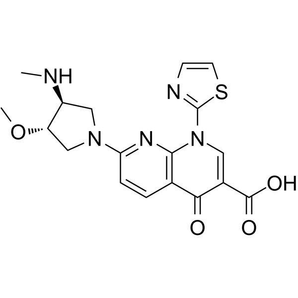
    Voreloxin (Synonyms: SNS-595; Vosaroxin; AG 7352)

    Voreloxin (SNS-595; Vosaroxin; AG 7352) 是一种新创的拓扑异构酶 II (topoisomerase II) 抑制剂,能够诱导 DNA 双链断裂,阻滞 G2 期,最终细胞凋亡。

    Voreloxin(Synonyms: SNS-595;  Vosaroxin;  AG 7352)

    Voreloxin Chemical Structure

    CAS No. : 175414-77-4

    规格 是否有货
    100 mg   询价  
    250 mg   询价  
    500 mg   询价  

    * Please select Quantity before adding items.

    Voreloxin 的其他形式现货产品:

    Voreloxin Hydrochloride

    生物活性

    Voreloxin (SNS-595; Vosaroxin; AG 7352) is a first-in-class topoisomerase II inhibitor that intercalates DNA and induces site-selective DNA DSB, G2 arrest, and apoptosis.

    IC50 & Target

    Topoisomerase II


    体外研究
    (In Vitro)

    Voreloxin is a first-in-class topoisomerase II poison and inhibitor that intercalates DNA and induces site-selective DNA DSB, G2 arrest, and apoptosis. Voreloxin (0.1-20 µM) inhibits topoisomerase II activity and induces site-selective DNA DSB in CCRF-CEM cells. Voreloxin (0.11, 0.33, 1, 3 µM) induces G2 arrest partially through topoisomerase II in A549 lung cancer cell line. Voreloxin cytotoxic activity requires DNA intercalation. However, Voreloxin (1-9 µM) does not generate significant levels of ROS[1]. Voreloxin has potent cytotoxic activity in AML cell lines MV4-11 and HL-60, with IC50s of 95 ± 8 nM and 884 ± 114 nM, respectively. Voreloxin in combination with cytarabine shows additive or synergistic activity in acute leukemia cell lines[2]. Voreloxin is active on the primary acute myeloid leukemia (AML) with a mean LD50 of 2.3 μM. The LD50 for voreloxin in myeloid cell lines NB4 and HL-60 is 0.59 μM ± 0.25 μM. Voreloxin causes accumulation of cells in the S and G2 phases of the cell cycle and acts on topoisomerase II[3].

    Shanghai Jinpan Biotech Co Ltd has not independently confirmed the accuracy of these methods. They are for reference only.

    体内研究
    (In Vivo)

    Voreloxin (20 mg/kg, i.v.) alone results in 80% reduction in bone marrow cellularity of CD-1 mice by administration one dose every 4 days repeated twice (q4d ×2). voreloxin at 10 mg/kg in combination with cytarabine causes ablation of the marrow, dilation of sinusoids, and infiltration of adipocytes in mice. Voreloxin (20 mg/kg, i.v.) combined with cytarabine causes a reversible decrease in myeloid and lymphoid cells in bone marrow and peripheral blood CD-1 mice. voreloxin (10 mg/kg, q4d ×2) and cytarabine in combination causes reversible neutropenia with a more modest impact on platelets CD-1 mice[2].

    Shanghai Jinpan Biotech Co Ltd has not independently confirmed the accuracy of these methods. They are for reference only.

    Clinical Trial

    分子量

    401.44

    Formula

    C18H19N5O4S

    CAS 号

    175414-77-4

    运输条件

    Room temperature in continental US; may vary elsewhere.

    储存方式

    Please store the product under the recommended conditions in the Certificate of Analysis.

    参考文献
    • [1]. Hotinski AK, et al. Vosaroxin is a novel topoisomerase-II inhibitor with efficacy in relapsed and refractory acute myeloid leukaemia. Expert Opin Pharmacother. 2015 Jun;16(9):1395-402.

      [2]. Scatena CD, et al. Voreloxin, a first-in-class anticancer quinolone derivative, acts synergistically with cytarabine in vitro and induces bone marrow aplasia in vivo. Cancer Chemother Pharmacol. 2010 Oct;66(5):881-8.

      [3]. Walsby EJ, et al. The topoisomerase II inhibitor voreloxin causes cell cycle arrest and apoptosis in myeloid leukemia cells and acts in synergy with cytarabine. Haematologica. 2011 Mar;96(3):393-9.

    Cell Assay
    [3]

    In vitro toxicity assays are performed on primary AML mononuclear cells over a 48 h period using a MTS cell proliferation assay. Lethal doses (LD50) are calculated. Cells are treated with voreloxin (31.25 nM to 4 μM) and Ara-C (62.5 nM to 8 μM) by serial dilution and incubated for 48 h in a final volume of 90 μL. Following the incubation, 20 μL of MTS reagent are added and the reaction is incubated for a further 4 h. The absorbance of the reaction after this time is read by spectrophotometry at 490 nm and the percentage of viable cells calculated relative to untreated control cells in the same assay. IC50 values are calculated using Calcusyn software[3].

    Shanghai Jinpan Biotech Co Ltd has not independently confirmed the accuracy of these methods. They are for reference only.

    Animal Administration
    [2]

    Animals are weighed, randomized by body weight, and assigned to the study groups before initiation of treatment. Voreloxin is administered intravenously (IV) at 10 or 20 mg/kg once on day zero and once on day four (q4d ×2). Cytarabine is administered subcutaneously (SC) at 20 or 60 mg/kg every 8 h on day zero and day four (tid q4d ×2). Tissues and blood are sampled on days 6, 8, and 12 from at least three and not greater than ten animals per treatment group. Femurs are placed in Streck Tissue Fixative solution, or in 10% formalin solution for 24-48 h followed by a 70% dehydrant (ethanol, isopropanol, methanol). Femurs are decalcified, paraffin embedded, and sectioned at Biopathology Labs. The four micron sections are stained with hematoxylin-eosin (H&E). H&E stained femurs are examined and percent cellularity of the bone marrow is determined. Digital photographs of representative femur sections are taken on a Leica DM2000 microscope using Image-Pro Plus v6.1 software[2].

    Shanghai Jinpan Biotech Co Ltd has not independently confirmed the accuracy of these methods. They are for reference only.

    参考文献
    • [1]. Hotinski AK, et al. Vosaroxin is a novel topoisomerase-II inhibitor with efficacy in relapsed and refractory acute myeloid leukaemia. Expert Opin Pharmacother. 2015 Jun;16(9):1395-402.

      [2]. Scatena CD, et al. Voreloxin, a first-in-class anticancer quinolone derivative, acts synergistically with cytarabine in vitro and induces bone marrow aplasia in vivo. Cancer Chemother Pharmacol. 2010 Oct;66(5):881-8.

      [3]. Walsby EJ, et al. The topoisomerase II inhibitor voreloxin causes cell cycle arrest and apoptosis in myeloid leukemia cells and acts in synergy with cytarabine. Haematologica. 2011 Mar;96(3):393-9.

    所有产品仅用作科学研究或药证申报,我们不为任何个人用途提供产品和服务
    Inolitazone(Synonyms: Efatutazone; CS-7017; RS5444)

    上海金畔生物科技有限公司为生命科学和医药研发人员提供生物活性分子抑制剂、激动剂、特异性抑制剂、化合物库、重组蛋白,专注于信号通路和疾病研究领域。

    Inolitazone (Synonyms: Efatutazone; CS-7017; RS5444)

    Inolitazone(RS5444; CS-7017)是新型PPARγ高亲和性激动剂,EC50为rosiglitazone的五十分之一,对于不表达PPARγ的RIE细胞无抑制性。

    Inolitazone(Synonyms: Efatutazone;  CS-7017;  RS5444)

    Inolitazone Chemical Structure

    CAS No. : 223132-37-4

    规格 是否有货
    100 mg   询价  
    250 mg   询价  
    500 mg   询价  

    * Please select Quantity before adding items.

    Inolitazone 的其他形式现货产品:

    Inolitazone dihydrochloride

    生物活性

    Inolitazone a novel high-affinity PPARγ agonist that is dependent upon PPARγ for its biological activity with IC50 of 0.8 nM for growth inhibition.

    IC50 & Target

    PPARγ


    体外研究
    (In Vitro)

    Inolitazone (RS5444) upregulates the cell cycle kinase inhibitor, p21 WAF1/CIP1. Silencing p21 WAF1/CIP1 rendered cells insensitive to Inolitazone. A 10 nM dose of Inolitazone activates PPARγ:RXRα-dependent transcription as demonstrated in a transient transfection assay utilizing a PPRE response element fused to a luciferase reporter gene (PPRE3-tk-luc). DRO cells are treated in culture with Inolitazone, Rosiglitazone, or Troglitazone at the indicated concentrations. DRO cells are transiently transfected with PPRE3-tk-luc to examine effective concentrations at which EC50 occurs. The EC50s are 1 nM (Inolitazone), 65 nM (Rosiglitazone) and 631 nM (Troglitazone). Similarly, the calculated inhibitory concentration at IC50 is 0.8 nM for Inolitazone, 75 nM for Rosiglitazone, and 1412 nM for Troglitazone. Inolitazone specifically activates PPARγ, but not PPARα or PPARδ. Exposure of 10 nM Inolitazone following transient transfection with the appropriate PPAR isoform (γ, α, or δ) and PPAR response element linked to a luciferase reporter in RIE rat small intestinal cell line, which does not express PPARs, yields increased luciferase activity only in the presence of PPARγ and PPRE3-tk-luc[1]. DRO cells are growth inhibited by 10 nM Inolitazone (RS5444) through a PPARγ-dependent mechanism[2].

    上海金畔生物科技有限公司 has not independently confirmed the accuracy of these methods. They are for reference only.

    体内研究
    (In Vivo)

    Inolitazone (RS5444) plus Paclitaxel demonstrate additive antiproliferative activity in cell culture and minimal ATC tumor growth. When Inolitazone is administered in the diet to athymic nude mice prior to DRO tumor cell implantation, tumor growth is inhibited in a dose responsive fashion. At the highest dose, 0.025% Inolitazone inhibits growth on day 32 by 94.4% as compared to that of control. In this treatment group, five of 10 animals do not develop demonstrable tumors. In the 0.0025% treatment group, tumor growth is inhibited by 62.3% compared to that of control on day 32 while the 0.00025% dose demonstrated no growth inhibitory activity as compared to control. Tumors is nest allowed to establish in the mouse and began 0.025% Inolitazone treatment of mice 1 week after DRO or ARO tumor cell implantation. Inolitazone treated animals demonstrate tumor growth inhibition of 68.9% in DRO tumors and 48.3% in ARO tumors as compared to that of their respective controls on day 35[1].

    上海金畔生物科技有限公司 has not independently confirmed the accuracy of these methods. They are for reference only.

    Clinical Trial

    分子量

    502.58

    Formula

    C27H26N4O4S

    CAS 号

    223132-37-4

    中文名称

    伊诺他酮二盐酸盐

    运输条件

    Room temperature in continental US; may vary elsewhere.

    储存方式

    Please store the product under the recommended conditions in the Certificate of Analysis.

    参考文献
    • [1]. Copland JA, et al. Novel high-affinity PPARgamma agonist alone and in combination with paclitaxel inhibits human anaplastic thyroid carcinoma tumor growth via p21WAF1/CIP1. Oncogene. 2006 Apr 13;25(16):2304-17.

      [2]. Marlow LA, et al. Reactivation of suppressed RhoB is a critical step for the inhibition of anaplastic thyroid cancer growth. Cancer Res. 2009 Feb 15;69(4):1536-44.

    Cell Assay
    [1]

    DRO90-1 (DRO) and ARO81 (ARO) cells are plated in 12-well culture plates in triplicate for each condition at an initial concentration of 2×10⁴ cells/well. After overnight incubation, cells are treated with either Inolitazone, Rosiglitazone, Troglitazone, GW9662, or Paclitaxel diluted in DMSO at concentrations indicated in figure legends. All cells receive identical volumes of DMSO and are exposed to each drug for 6 days with medium and drug changed every 48 h. After 6 days, cells are washed with PBS, trypsinized and counted by Beckman Coulter Counter[1].

    上海金畔生物科技有限公司 has not independently confirmed the accuracy of these methods. They are for reference only.

    Animal Administration
    [1]

    Mice[1]
    Suspensions of 1×10⁶/0.1 mL DRO or ARO cells in RPMI medium are injected subcutaneously in one flank of 3-4 week athymic female nude mice. Mice are changed to specialized diets either 1 week prior or 1 week after tumor implantation and randomly assigned to experimental or control groups with 10 mice per group. Diets consisted either placebo, 0.00025%, 0.0025%, or 0.025% Inolitazone formulated into the diet. Mice weighed between 20-25 g and consume on average 4 g of food per day. For combinatorial studies either placebo, 10 mg/kg or 15 mg/kg paclitaxel is injected i.p. twice weekly. Tumors are measured every 3-4 days for 35 days with calipers and tumor volumes are calculated by the formula: 0.5236(a×b×c), where a is the shortest diameter, b is the diameter perpendicular to a and c is the diameter height.

    上海金畔生物科技有限公司 has not independently confirmed the accuracy of these methods. They are for reference only.

    参考文献
    • [1]. Copland JA, et al. Novel high-affinity PPARgamma agonist alone and in combination with paclitaxel inhibits human anaplastic thyroid carcinoma tumor growth via p21WAF1/CIP1. Oncogene. 2006 Apr 13;25(16):2304-17.

      [2]. Marlow LA, et al. Reactivation of suppressed RhoB is a critical step for the inhibition of anaplastic thyroid cancer growth. Cancer Res. 2009 Feb 15;69(4):1536-44.

    所有产品仅用作科学研究或药证申报,我们不为任何个人用途提供产品和服务

    Inolitazone(Synonyms: Efatutazone; CS-7017; RS5444)

    上海金畔生物科技有限公司为生命科学和医药研发人员提供生物活性分子抑制剂、激动剂、特异性抑制剂、化合物库、重组蛋白,专注于信号通路和疾病研究领域。

    Inolitazone (Synonyms: Efatutazone; CS-7017; RS5444)

    Inolitazone(RS5444; CS-7017)是新型PPARγ高亲和性激动剂,EC50为rosiglitazone的五十分之一,对于不表达PPARγ的RIE细胞无抑制性。

    Inolitazone(Synonyms: Efatutazone;  CS-7017;  RS5444)

    Inolitazone Chemical Structure

    CAS No. : 223132-37-4

    规格 是否有货
    100 mg   询价  
    250 mg   询价  
    500 mg   询价  

    * Please select Quantity before adding items.

    Inolitazone 的其他形式现货产品:

    Inolitazone dihydrochloride

    生物活性

    Inolitazone a novel high-affinity PPARγ agonist that is dependent upon PPARγ for its biological activity with IC50 of 0.8 nM for growth inhibition.

    IC50 & Target

    PPARγ


    体外研究
    (In Vitro)

    Inolitazone (RS5444) upregulates the cell cycle kinase inhibitor, p21 WAF1/CIP1. Silencing p21 WAF1/CIP1 rendered cells insensitive to Inolitazone. A 10 nM dose of Inolitazone activates PPARγ:RXRα-dependent transcription as demonstrated in a transient transfection assay utilizing a PPRE response element fused to a luciferase reporter gene (PPRE3-tk-luc). DRO cells are treated in culture with Inolitazone, Rosiglitazone, or Troglitazone at the indicated concentrations. DRO cells are transiently transfected with PPRE3-tk-luc to examine effective concentrations at which EC50 occurs. The EC50s are 1 nM (Inolitazone), 65 nM (Rosiglitazone) and 631 nM (Troglitazone). Similarly, the calculated inhibitory concentration at IC50 is 0.8 nM for Inolitazone, 75 nM for Rosiglitazone, and 1412 nM for Troglitazone. Inolitazone specifically activates PPARγ, but not PPARα or PPARδ. Exposure of 10 nM Inolitazone following transient transfection with the appropriate PPAR isoform (γ, α, or δ) and PPAR response element linked to a luciferase reporter in RIE rat small intestinal cell line, which does not express PPARs, yields increased luciferase activity only in the presence of PPARγ and PPRE3-tk-luc[1]. DRO cells are growth inhibited by 10 nM Inolitazone (RS5444) through a PPARγ-dependent mechanism[2].

    Shanghai Jinpan Biotech Co Ltd has not independently confirmed the accuracy of these methods. They are for reference only.

    体内研究
    (In Vivo)

    Inolitazone (RS5444) plus Paclitaxel demonstrate additive antiproliferative activity in cell culture and minimal ATC tumor growth. When Inolitazone is administered in the diet to athymic nude mice prior to DRO tumor cell implantation, tumor growth is inhibited in a dose responsive fashion. At the highest dose, 0.025% Inolitazone inhibits growth on day 32 by 94.4% as compared to that of control. In this treatment group, five of 10 animals do not develop demonstrable tumors. In the 0.0025% treatment group, tumor growth is inhibited by 62.3% compared to that of control on day 32 while the 0.00025% dose demonstrated no growth inhibitory activity as compared to control. Tumors is nest allowed to establish in the mouse and began 0.025% Inolitazone treatment of mice 1 week after DRO or ARO tumor cell implantation. Inolitazone treated animals demonstrate tumor growth inhibition of 68.9% in DRO tumors and 48.3% in ARO tumors as compared to that of their respective controls on day 35[1].

    Shanghai Jinpan Biotech Co Ltd has not independently confirmed the accuracy of these methods. They are for reference only.

    Clinical Trial

    分子量

    502.58

    Formula

    C27H26N4O4S

    CAS 号

    223132-37-4

    中文名称

    伊诺他酮二盐酸盐

    运输条件

    Room temperature in continental US; may vary elsewhere.

    储存方式

    Please store the product under the recommended conditions in the Certificate of Analysis.

    参考文献
    • [1]. Copland JA, et al. Novel high-affinity PPARgamma agonist alone and in combination with paclitaxel inhibits human anaplastic thyroid carcinoma tumor growth via p21WAF1/CIP1. Oncogene. 2006 Apr 13;25(16):2304-17.

      [2]. Marlow LA, et al. Reactivation of suppressed RhoB is a critical step for the inhibition of anaplastic thyroid cancer growth. Cancer Res. 2009 Feb 15;69(4):1536-44.

    Cell Assay
    [1]

    DRO90-1 (DRO) and ARO81 (ARO) cells are plated in 12-well culture plates in triplicate for each condition at an initial concentration of 2×10⁴ cells/well. After overnight incubation, cells are treated with either Inolitazone, Rosiglitazone, Troglitazone, GW9662, or Paclitaxel diluted in DMSO at concentrations indicated in figure legends. All cells receive identical volumes of DMSO and are exposed to each drug for 6 days with medium and drug changed every 48 h. After 6 days, cells are washed with PBS, trypsinized and counted by Beckman Coulter Counter[1].

    Shanghai Jinpan Biotech Co Ltd has not independently confirmed the accuracy of these methods. They are for reference only.

    Animal Administration
    [1]

    Mice[1]
    Suspensions of 1×10⁶/0.1 mL DRO or ARO cells in RPMI medium are injected subcutaneously in one flank of 3-4 week athymic female nude mice. Mice are changed to specialized diets either 1 week prior or 1 week after tumor implantation and randomly assigned to experimental or control groups with 10 mice per group. Diets consisted either placebo, 0.00025%, 0.0025%, or 0.025% Inolitazone formulated into the diet. Mice weighed between 20-25 g and consume on average 4 g of food per day. For combinatorial studies either placebo, 10 mg/kg or 15 mg/kg paclitaxel is injected i.p. twice weekly. Tumors are measured every 3-4 days for 35 days with calipers and tumor volumes are calculated by the formula: 0.5236(a×b×c), where a is the shortest diameter, b is the diameter perpendicular to a and c is the diameter height.

    Shanghai Jinpan Biotech Co Ltd has not independently confirmed the accuracy of these methods. They are for reference only.

    参考文献
    • [1]. Copland JA, et al. Novel high-affinity PPARgamma agonist alone and in combination with paclitaxel inhibits human anaplastic thyroid carcinoma tumor growth via p21WAF1/CIP1. Oncogene. 2006 Apr 13;25(16):2304-17.

      [2]. Marlow LA, et al. Reactivation of suppressed RhoB is a critical step for the inhibition of anaplastic thyroid cancer growth. Cancer Res. 2009 Feb 15;69(4):1536-44.

    所有产品仅用作科学研究或药证申报,我们不为任何个人用途提供产品和服务

    ZM323881

    上海金畔生物科技有限公司为生命科学和医药研发人员提供生物活性分子抑制剂、激动剂、特异性抑制剂、化合物库、重组蛋白,专注于信号通路和疾病研究领域。

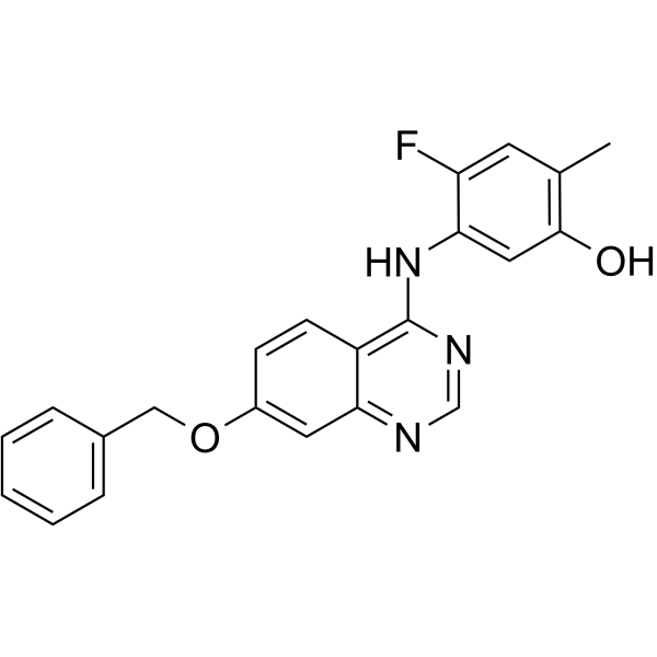
    ZM323881 

    ZM323881是有效,选择性的 VEGFR2 抑制剂, IC50 值小于2 nM。

    ZM323881

    ZM323881 Chemical Structure

    CAS No. : 193001-14-8

    规格 是否有货
    100 mg   询价  
    250 mg   询价  
    500 mg   询价  

    * Please select Quantity before adding items.

    ZM323881 的其他形式现货产品:

    ZM323881 hydrochloride

    生物活性

    ZM323881 is a potent and selective VEGFR2 inhibitor with an IC50 of less than 2 nM.

    IC50 & Target[1]

    VEGFR2

    2 nM (IC50)

    体外研究
    (In Vitro)

    ZM323881 is an anilinoquinazoline that potently inhibits VEGFR2 (KDR) tyrosine kinase activity anddemonstrates excellent selectivity versus other receptor tyrosine kinases, including PDGFRβ, FGFR1, EGFR and erbB2 (IC50>50 μM). ZM323881 inhibits VEGF-A-induced endothelial cell proliferation(IC50=8 nM) and VEGFR2 tyrosine phosphorylation[1]. ZM323881 inhibits activation of VEGFR-2, but not of VEGFR-1, epidermal growth factor receptor (EGFR), platelet-derived growth factor receptor (PDGFR), or hepatocyte growth factor (HGF) receptor. In HAECs, ZM323881 completely inhibits VEGF-induced ERK phosphorylation at 1 μM[2].

    Shanghai Jinpan Biotech Co Ltd has not independently confirmed the accuracy of these methods. They are for reference only.

    分子量

    375.40

    Formula

    C22H18FN3O2

    CAS 号

    193001-14-8

    运输条件

    Room temperature in continental US; may vary elsewhere.

    储存方式

    Please store the product under the recommended conditions in the Certificate of Analysis.

    参考文献
    • [1]. Whittles CE, et al. ZM323881, a novel inhibitor of vascular endothelial growth factor-receptor-2 tyrosine kinase activity. Microcirculation. 2002 Dec;9(6):513-22.

      [2]. Endo A, et al. Selective inhibition of vascular endothelial growth factor receptor-2 (VEGFR-2) identifies a central role for VEGFR-2 in human aortic endothelial cell responses to VEGF. J Recept Signal Transduct Res. 2003;23(2-3):239-54.

    Kinase Assay
    [1]

    Compounds (ZM323881) are incubated (20 minutes, room temperature) with enzyme in an N-2-hydroxyethylpiperazine-N’-2-ethanesulphonate (HEPES) (pH 7.5) buffered solution containing 10 mM MnCl2 and 2 μM ATP, in96-well plates coated with a poly(Glu, Ala, Tyr) 6:3:1 random copolymer substrate. Phosphorylated tyrosine is then detected bysequential incubation with mouse IgG anti-phosphotyrosine antibody a horseradish peroxidase(HRP)-linked sheep anti-mouse Ig antibody and 2,2′-azino-bis(3-ethylbenzthiazoline-6-sulphonic acid). IC50 data are interpolated by nonlin-ear regression[1].

    Shanghai Jinpan Biotech Co Ltd has not independently confirmed the accuracy of these methods. They are for reference only.

    Cell Assay
    [1]

    HUVEC cells isolated from umbilical cords are plated (at passage 2–8) in 96-wellplates (1000 cells/well) and dosed with ZM323881±VEGF-A (3 ng/mL), EGF (3 ng/mL), or basicfibroblast growth factor (bFGF, 0.3 ng/mL). The cultures are then incubated for 4 days. On day 4, the cultures are pulsed with 1 μCi/well of 3H-thymidine and reincubated for 4 hours. The cells are then harvested and assayed for the incorporation of tritium by using a beta-counter. IC50 data are interpolated[1].

    Shanghai Jinpan Biotech Co Ltd has not independently confirmed the accuracy of these methods. They are for reference only.

    参考文献
    • [1]. Whittles CE, et al. ZM323881, a novel inhibitor of vascular endothelial growth factor-receptor-2 tyrosine kinase activity. Microcirculation. 2002 Dec;9(6):513-22.

      [2]. Endo A, et al. Selective inhibition of vascular endothelial growth factor receptor-2 (VEGFR-2) identifies a central role for VEGFR-2 in human aortic endothelial cell responses to VEGF. J Recept Signal Transduct Res. 2003;23(2-3):239-54.

    所有产品仅用作科学研究或药证申报,我们不为任何个人用途提供产品和服务

    Voreloxin(Synonyms: SNS-595; Vosaroxin; AG 7352)

    上海金畔生物科技有限公司为生命科学和医药研发人员提供生物活性分子抑制剂、激动剂、特异性抑制剂、化合物库、重组蛋白,专注于信号通路和疾病研究领域。

    Voreloxin (Synonyms: SNS-595; Vosaroxin; AG 7352)

    Voreloxin (SNS-595; Vosaroxin; AG 7352) 是一种新创的拓扑异构酶 II (topoisomerase II) 抑制剂,能够诱导 DNA 双链断裂,阻滞 G2 期,最终细胞凋亡。

    Voreloxin(Synonyms: SNS-595;  Vosaroxin;  AG 7352)

    Voreloxin Chemical Structure

    CAS No. : 175414-77-4

    规格 是否有货
    100 mg   询价  
    250 mg   询价  
    500 mg   询价  

    * Please select Quantity before adding items.

    Voreloxin 的其他形式现货产品:

    Voreloxin Hydrochloride

    生物活性

    Voreloxin (SNS-595; Vosaroxin; AG 7352) is a first-in-class topoisomerase II inhibitor that intercalates DNA and induces site-selective DNA DSB, G2 arrest, and apoptosis.

    IC50 & Target

    Topoisomerase II


    体外研究
    (In Vitro)

    Voreloxin is a first-in-class topoisomerase II poison and inhibitor that intercalates DNA and induces site-selective DNA DSB, G2 arrest, and apoptosis. Voreloxin (0.1-20 µM) inhibits topoisomerase II activity and induces site-selective DNA DSB in CCRF-CEM cells. Voreloxin (0.11, 0.33, 1, 3 µM) induces G2 arrest partially through topoisomerase II in A549 lung cancer cell line. Voreloxin cytotoxic activity requires DNA intercalation. However, Voreloxin (1-9 µM) does not generate significant levels of ROS[1]. Voreloxin has potent cytotoxic activity in AML cell lines MV4-11 and HL-60, with IC50s of 95 ± 8 nM and 884 ± 114 nM, respectively. Voreloxin in combination with cytarabine shows additive or synergistic activity in acute leukemia cell lines[2]. Voreloxin is active on the primary acute myeloid leukemia (AML) with a mean LD50 of 2.3 μM. The LD50 for voreloxin in myeloid cell lines NB4 and HL-60 is 0.59 μM ± 0.25 μM. Voreloxin causes accumulation of cells in the S and G2 phases of the cell cycle and acts on topoisomerase II[3].

    上海金畔生物科技有限公司 has not independently confirmed the accuracy of these methods. They are for reference only.

    体内研究
    (In Vivo)

    Voreloxin (20 mg/kg, i.v.) alone results in 80% reduction in bone marrow cellularity of CD-1 mice by administration one dose every 4 days repeated twice (q4d ×2). voreloxin at 10 mg/kg in combination with cytarabine causes ablation of the marrow, dilation of sinusoids, and infiltration of adipocytes in mice. Voreloxin (20 mg/kg, i.v.) combined with cytarabine causes a reversible decrease in myeloid and lymphoid cells in bone marrow and peripheral blood CD-1 mice. voreloxin (10 mg/kg, q4d ×2) and cytarabine in combination causes reversible neutropenia with a more modest impact on platelets CD-1 mice[2].

    上海金畔生物科技有限公司 has not independently confirmed the accuracy of these methods. They are for reference only.

    Clinical Trial

    分子量

    401.44

    Formula

    C18H19N5O4S

    CAS 号

    175414-77-4

    运输条件

    Room temperature in continental US; may vary elsewhere.

    储存方式

    Please store the product under the recommended conditions in the Certificate of Analysis.

    参考文献
    • [1]. Hotinski AK, et al. Vosaroxin is a novel topoisomerase-II inhibitor with efficacy in relapsed and refractory acute myeloid leukaemia. Expert Opin Pharmacother. 2015 Jun;16(9):1395-402.

      [2]. Scatena CD, et al. Voreloxin, a first-in-class anticancer quinolone derivative, acts synergistically with cytarabine in vitro and induces bone marrow aplasia in vivo. Cancer Chemother Pharmacol. 2010 Oct;66(5):881-8.

      [3]. Walsby EJ, et al. The topoisomerase II inhibitor voreloxin causes cell cycle arrest and apoptosis in myeloid leukemia cells and acts in synergy with cytarabine. Haematologica. 2011 Mar;96(3):393-9.

    Cell Assay
    [3]

    In vitro toxicity assays are performed on primary AML mononuclear cells over a 48 h period using a MTS cell proliferation assay. Lethal doses (LD50) are calculated. Cells are treated with voreloxin (31.25 nM to 4 μM) and Ara-C (62.5 nM to 8 μM) by serial dilution and incubated for 48 h in a final volume of 90 μL. Following the incubation, 20 μL of MTS reagent are added and the reaction is incubated for a further 4 h. The absorbance of the reaction after this time is read by spectrophotometry at 490 nm and the percentage of viable cells calculated relative to untreated control cells in the same assay. IC50 values are calculated using Calcusyn software[3].

    上海金畔生物科技有限公司 has not independently confirmed the accuracy of these methods. They are for reference only.

    Animal Administration
    [2]

    Animals are weighed, randomized by body weight, and assigned to the study groups before initiation of treatment. Voreloxin is administered intravenously (IV) at 10 or 20 mg/kg once on day zero and once on day four (q4d ×2). Cytarabine is administered subcutaneously (SC) at 20 or 60 mg/kg every 8 h on day zero and day four (tid q4d ×2). Tissues and blood are sampled on days 6, 8, and 12 from at least three and not greater than ten animals per treatment group. Femurs are placed in Streck Tissue Fixative solution, or in 10% formalin solution for 24-48 h followed by a 70% dehydrant (ethanol, isopropanol, methanol). Femurs are decalcified, paraffin embedded, and sectioned at Biopathology Labs. The four micron sections are stained with hematoxylin-eosin (H&E). H&E stained femurs are examined and percent cellularity of the bone marrow is determined. Digital photographs of representative femur sections are taken on a Leica DM2000 microscope using Image-Pro Plus v6.1 software[2].

    上海金畔生物科技有限公司 has not independently confirmed the accuracy of these methods. They are for reference only.

    参考文献
    • [1]. Hotinski AK, et al. Vosaroxin is a novel topoisomerase-II inhibitor with efficacy in relapsed and refractory acute myeloid leukaemia. Expert Opin Pharmacother. 2015 Jun;16(9):1395-402.

      [2]. Scatena CD, et al. Voreloxin, a first-in-class anticancer quinolone derivative, acts synergistically with cytarabine in vitro and induces bone marrow aplasia in vivo. Cancer Chemother Pharmacol. 2010 Oct;66(5):881-8.

      [3]. Walsby EJ, et al. The topoisomerase II inhibitor voreloxin causes cell cycle arrest and apoptosis in myeloid leukemia cells and acts in synergy with cytarabine. Haematologica. 2011 Mar;96(3):393-9.

    所有产品仅用作科学研究或药证申报,我们不为任何个人用途提供产品和服务

    多肽定制TNF-α (10-36) (human) 编码

    上海金畔生物科技有限公司可以定制不同序列多肽,可以访问官网了解更多产品信息。

    名称 TNF-α (10-36) (human)
    编码
    别名 TNF-α (10-36) (human)
    纯度 80%,90%,95%,98%,99%
    重量 1mg,5mg,10mg,50mg,100mg,1g
    序列(单字母缩写) DKPVAHVVANPQAEGQLQWLNRRANAL-OH
    序列(三字母缩写) Asp-Lys-Pro-Val-Ala-His-Val-Val-Ala-Asn-Pro-Gln-Ala-Glu-Gly-Gln-Leu-Gln-Trp-Leu-Asn-Arg-Arg-Ala-Asn-Ala-Leu
    基本描述
    溶解度
    分子量 0
    化学式
    存储条件 Store at -20°C. Keep tightly closed. Store in a cool dry place.
    注释
    Documents TNF-α (10-36) (human)          编码
    Figures TNF-α (10-36) (human)          编码
    Reference
    C端
    N端
    化学桥

    VM-80垂直旋转混合仪

    【简单介绍】

    VM-80垂直旋转混合仪提供高效且温和的混合,保持生物样品处于悬浮状态,如血液混合。适用于预防血液凝固、乳胶诊断、免疫沉淀等相似应用。

    【详细说明】

    VM-80垂直旋转混合仪

    产品简述:

      MS-20/MS-40数字式顶置搅拌器提供高效且温和的混合,保持生物样品处于悬浮状态,如血液混合。适用于预防血液凝固、乳胶诊断、免疫沉淀等相似应用。


    产品特点: 

    1、外形精巧,构造坚固,操作简单,LED显示屏显示时间转速。

    2、无刷直流电机性能稳定,低噪音,转速稳定,使用寿命长。 

    3、360°垂直循环旋转,运转平稳。

    4、可更换不同平台,兼容使用不同尺寸的试管。


    VM-80垂直旋转混合仪

    技术参数

    产品型号

    VM-80旋转混匀仪

    电压

    100-240VAC   50/60Hz

    电机输入功率

    16W

    电机输出功率

    10W

    速度范围

    10-80rpm

    运转方式

    圆周运转

    外观尺寸(长×宽×高)(mm)

    470×190 ×160

    重量

    2.8kg


    可更换夹具

    型号

    描述

    MV80-A

    1.5ml  x 64

    MV80-B

    15ml  x22

    MV80-C

    50ml  x16


    Voreloxin(Synonyms: SNS-595; Vosaroxin; AG 7352)

    上海金畔生物科技有限公司为生命科学和医药研发人员提供生物活性分子抑制剂、激动剂、特异性抑制剂、化合物库、重组蛋白,专注于信号通路和疾病研究领域。

    Voreloxin (Synonyms: SNS-595; Vosaroxin; AG 7352)

    Voreloxin (SNS-595; Vosaroxin; AG 7352) 是一种新创的拓扑异构酶 II (topoisomerase II) 抑制剂,能够诱导 DNA 双链断裂,阻滞 G2 期,最终细胞凋亡。

    Voreloxin(Synonyms: SNS-595;  Vosaroxin;  AG 7352)

    Voreloxin Chemical Structure

    CAS No. : 175414-77-4

    规格 是否有货
    100 mg   询价  
    250 mg   询价  
    500 mg   询价  

    * Please select Quantity before adding items.

    Voreloxin 的其他形式现货产品:

    Voreloxin Hydrochloride

    生物活性

    Voreloxin (SNS-595; Vosaroxin; AG 7352) is a first-in-class topoisomerase II inhibitor that intercalates DNA and induces site-selective DNA DSB, G2 arrest, and apoptosis.

    IC50 & Target

    Topoisomerase II


    体外研究
    (In Vitro)

    Voreloxin is a first-in-class topoisomerase II poison and inhibitor that intercalates DNA and induces site-selective DNA DSB, G2 arrest, and apoptosis. Voreloxin (0.1-20 µM) inhibits topoisomerase II activity and induces site-selective DNA DSB in CCRF-CEM cells. Voreloxin (0.11, 0.33, 1, 3 µM) induces G2 arrest partially through topoisomerase II in A549 lung cancer cell line. Voreloxin cytotoxic activity requires DNA intercalation. However, Voreloxin (1-9 µM) does not generate significant levels of ROS[1]. Voreloxin has potent cytotoxic activity in AML cell lines MV4-11 and HL-60, with IC50s of 95 ± 8 nM and 884 ± 114 nM, respectively. Voreloxin in combination with cytarabine shows additive or synergistic activity in acute leukemia cell lines[2]. Voreloxin is active on the primary acute myeloid leukemia (AML) with a mean LD50 of 2.3 μM. The LD50 for voreloxin in myeloid cell lines NB4 and HL-60 is 0.59 μM ± 0.25 μM. Voreloxin causes accumulation of cells in the S and G2 phases of the cell cycle and acts on topoisomerase II[3].

    Shanghai Jinpan Biotech Co Ltd has not independently confirmed the accuracy of these methods. They are for reference only.

    体内研究
    (In Vivo)

    Voreloxin (20 mg/kg, i.v.) alone results in 80% reduction in bone marrow cellularity of CD-1 mice by administration one dose every 4 days repeated twice (q4d ×2). voreloxin at 10 mg/kg in combination with cytarabine causes ablation of the marrow, dilation of sinusoids, and infiltration of adipocytes in mice. Voreloxin (20 mg/kg, i.v.) combined with cytarabine causes a reversible decrease in myeloid and lymphoid cells in bone marrow and peripheral blood CD-1 mice. voreloxin (10 mg/kg, q4d ×2) and cytarabine in combination causes reversible neutropenia with a more modest impact on platelets CD-1 mice[2].

    Shanghai Jinpan Biotech Co Ltd has not independently confirmed the accuracy of these methods. They are for reference only.

    Clinical Trial

    分子量

    401.44

    Formula

    C18H19N5O4S

    CAS 号

    175414-77-4

    运输条件

    Room temperature in continental US; may vary elsewhere.

    储存方式

    Please store the product under the recommended conditions in the Certificate of Analysis.

    参考文献
    • [1]. Hotinski AK, et al. Vosaroxin is a novel topoisomerase-II inhibitor with efficacy in relapsed and refractory acute myeloid leukaemia. Expert Opin Pharmacother. 2015 Jun;16(9):1395-402.

      [2]. Scatena CD, et al. Voreloxin, a first-in-class anticancer quinolone derivative, acts synergistically with cytarabine in vitro and induces bone marrow aplasia in vivo. Cancer Chemother Pharmacol. 2010 Oct;66(5):881-8.

      [3]. Walsby EJ, et al. The topoisomerase II inhibitor voreloxin causes cell cycle arrest and apoptosis in myeloid leukemia cells and acts in synergy with cytarabine. Haematologica. 2011 Mar;96(3):393-9.

    Cell Assay
    [3]

    In vitro toxicity assays are performed on primary AML mononuclear cells over a 48 h period using a MTS cell proliferation assay. Lethal doses (LD50) are calculated. Cells are treated with voreloxin (31.25 nM to 4 μM) and Ara-C (62.5 nM to 8 μM) by serial dilution and incubated for 48 h in a final volume of 90 μL. Following the incubation, 20 μL of MTS reagent are added and the reaction is incubated for a further 4 h. The absorbance of the reaction after this time is read by spectrophotometry at 490 nm and the percentage of viable cells calculated relative to untreated control cells in the same assay. IC50 values are calculated using Calcusyn software[3].

    Shanghai Jinpan Biotech Co Ltd has not independently confirmed the accuracy of these methods. They are for reference only.

    Animal Administration
    [2]

    Animals are weighed, randomized by body weight, and assigned to the study groups before initiation of treatment. Voreloxin is administered intravenously (IV) at 10 or 20 mg/kg once on day zero and once on day four (q4d ×2). Cytarabine is administered subcutaneously (SC) at 20 or 60 mg/kg every 8 h on day zero and day four (tid q4d ×2). Tissues and blood are sampled on days 6, 8, and 12 from at least three and not greater than ten animals per treatment group. Femurs are placed in Streck Tissue Fixative solution, or in 10% formalin solution for 24-48 h followed by a 70% dehydrant (ethanol, isopropanol, methanol). Femurs are decalcified, paraffin embedded, and sectioned at Biopathology Labs. The four micron sections are stained with hematoxylin-eosin (H&E). H&E stained femurs are examined and percent cellularity of the bone marrow is determined. Digital photographs of representative femur sections are taken on a Leica DM2000 microscope using Image-Pro Plus v6.1 software[2].

    Shanghai Jinpan Biotech Co Ltd has not independently confirmed the accuracy of these methods. They are for reference only.

    参考文献
    • [1]. Hotinski AK, et al. Vosaroxin is a novel topoisomerase-II inhibitor with efficacy in relapsed and refractory acute myeloid leukaemia. Expert Opin Pharmacother. 2015 Jun;16(9):1395-402.

      [2]. Scatena CD, et al. Voreloxin, a first-in-class anticancer quinolone derivative, acts synergistically with cytarabine in vitro and induces bone marrow aplasia in vivo. Cancer Chemother Pharmacol. 2010 Oct;66(5):881-8.

      [3]. Walsby EJ, et al. The topoisomerase II inhibitor voreloxin causes cell cycle arrest and apoptosis in myeloid leukemia cells and acts in synergy with cytarabine. Haematologica. 2011 Mar;96(3):393-9.

    所有产品仅用作科学研究或药证申报,我们不为任何个人用途提供产品和服务

    郑州长城科工贸GRS玻璃反应釜GRS-50

    郑州长城科工贸GRS玻璃反应釜GRS-50

  • 品牌 长城科工贸|Greatwall
  • 型号 GRS-50
  • 商品详情

    专利号:201630293823.8

    用途特点:

    ●框架为不锈钢材质,防腐蚀。

    ●配置316材质接料托盘,可回收物料,防止污染环境。

    ●高硼硅玻璃材质,具有优良的物理、化学性能。

    ●可在高温(200℃)至低温(-80℃)的范围内使用。

    ●可在常压及负压条件下工作,真空度可达0.095MPa。

    ●釜内可承受压力范围:-0.1MPa~0MPa。

    ●特氟隆+FV橡胶材质的旋塞或放料阀;外加四氟乙烯保护层的“O”型圈。

    型号

    GRS-20

    GRS-20EX

    功率(减速比:3)(W)

    90

    180

    搅拌转速(rpm)

    50~500

    最大扭矩(N*m)

    0.6

    物料容积(L)

    20

    夹套容积(L)

    6

    电源

    220-240V~,50/60Hz

    放料阀离地高(mm)

    550

    整机高度(mm)

    2135


    型号

    GRS-50

    GRS-50EX

    功率(减速比:3)(W)

    140

    180

    搅拌转速(rpm)

    50~500

    最大扭矩(N*m)

    0.95

    物料容积(L)

    50

    夹套容积(L)

    16

    电源

    220-240V~,50/60Hz

    放料阀离地高(mm)

    550

    整机高度(mm)

    2650

  • CAL-130

    上海金畔生物科技有限公司为生命科学和医药研发人员提供生物活性分子抑制剂、激动剂、特异性抑制剂、化合物库、重组蛋白,专注于信号通路和疾病研究领域。

    CAL-130 

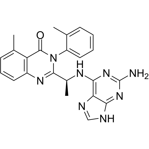
    CAL-130 是一种 PI3KδPI3Kγ 抑制剂,IC50 分别为 1.3 和 6.1 nM。

    CAL-130

    CAL-130 Chemical Structure

    CAS No. : 1431697-74-3

    规格 是否有货
    100 mg   询价  
    250 mg   询价  
    500 mg   询价  

    * Please select Quantity before adding items.

    CAL-130 的其他形式现货产品:

    CAL-130 Hydrochloride

    生物活性

    CAL-130 is a PI3Kδ and PI3Kγ inhibitor with IC50s of 1.3 and 6.1 nM, respectively.

    IC50 & Target[1]

    p110δ

    1.3 nM (IC50)

    p110γ

    6.1 nM (IC50)

    p110β

    56 nM (IC50)

    p110α

    115 nM (IC50)

    体外研究
    (In Vitro)

    CAL-130 preferentially inhibits the function of both p110γ and p110δ catalytic domains. IC50 values of CAL-130 are 1.3 and 6.1 nM for p110δ and p110γ, respectively, as compared to 115 and 56 nM for p110α and p110β. CAL-130 does not inhibit additional intracellular signaling pathways (i.e., p38 MAPK or insulin receptor tyrosine kinase) that are critical for general cell function and survival[1].

    Shanghai Jinpan Biotech Co Ltd has not independently confirmed the accuracy of these methods. They are for reference only.

    体内研究
    (In Vivo)

    The clinical significance of interfering with the combined activities of PI3Kγ and PI3Kδ is determined by administering CAL-130 to Lck/Ptenfl/fl mice with established T cell acute lymphoblastic leukemia (T-ALL). Candidate animals for survival studies are ill appearing, have a white blood cell (WBC) count above 45,000 μL-1, evidence of blasts on peripheral smear, and a majority of circulation cells (>75%) staining double positive for Thy1.2 and Ki-67. Mice receive an oral dose (10 mg/kg) of CAL-130 every 8 hr for a period of 7 days and are then followed until moribund. Despite the limited duration of therapy, CAL-130 is highly effective in extending the median survival for treated animals to 45 days as compared 7.5 days for the control group[1].

    Shanghai Jinpan Biotech Co Ltd has not independently confirmed the accuracy of these methods. They are for reference only.

    分子量

    426.47

    Formula

    C23H22N8O

    CAS 号

    1431697-74-3

    运输条件

    Room temperature in continental US; may vary elsewhere.

    储存方式

    Please store the product under the recommended conditions in the Certificate of Analysis.

    参考文献
    • [1]. Subramaniam Prem S, et al. Targeting nonclassical oncogenes for therapy in T-ALL. Cancer cell (2012), 21(4), 459-72.

    Kinase Assay
    [1]

    IC50 values for CAL-130 inhibition of PI3K isoforms are determined in ex vivo PI3 kinase assays using recombinant PI3K. A ten-point kinase inhibitory profile is determined with ATP at a concentration consistent with the KM for each enzyme[1].

    Shanghai Jinpan Biotech Co Ltd has not independently confirmed the accuracy of these methods. They are for reference only.

    Cell Assay
    [1]

    Cell proliferation of CCRF-CEM cells or shRNA-transfected CCRF-CEM cells, in presence or absence of CAL-130 (1, 2.5 and 5 μM), is followed by cell counting of samples in triplicate using a hemocytometer and trypan blue. For apoptosis determinations of untransfected or shRNA-transfected CCRF-CEMs, cells are stained with APC-conjugated Annexin-V in Annexin Binding Buffer and analyzed by flow cytometry. For primary T-ALL samples, cell viability is assessed using the BD Cell Viability kit coupled with the use of fluorescentcounting beads. For this, cells are plated with MS5-DL1 stroma cells, and after 72 hr following CAL-130 treatment, cells are harvested and stained with an APC-conjugated antihuman CD45 followed by a staining with the aforementioned kit[1].

    Shanghai Jinpan Biotech Co Ltd has not independently confirmed the accuracy of these methods. They are for reference only.

    Animal Administration
    [1]

    Mice[1]
    For subcutaneous xenograft experiments, luminescent CCRF-CEM (CEMluc) cells are generated by lentiviral infection with FUW-luc and selection with Neomycin. Luciferase expression is verified with the Dual-Luciferase Reporter Assay kit. 2.5×10⁶ CEM-luc cells embedded in Matrigel are injected in the flank of NOD.Cg-Prkdcscid Il2rgtm1Wjl/Sz mice. After 1 week, mice are treated by oral gavage with vehicle (0.5% methyl cellulose, 0.1% Tween 80), or CAL-130 (10 mg/kg) every 8 hr daily for 4 days, and then tumors are imaged as follows: mice anesthetized by isoflurane inhalation are injected intraperitoneally with D-luciferin (50 mg/kg). Photonic emission is imaged with the in vivo imaging system. Tumor bioluminescence is quantified by integrating the photonic flux (photons per second) through a region encircling each tumor using the Living Image software package. Administration of D-luciferin and detection of tumor bioluminescence in Lck/Ptenfl/fl/Gt(ROSA)26Sortm1(Luc)Kael/J mice are performed in a similar manner.

    Shanghai Jinpan Biotech Co Ltd has not independently confirmed the accuracy of these methods. They are for reference only.

    参考文献
    • [1]. Subramaniam Prem S, et al. Targeting nonclassical oncogenes for therapy in T-ALL. Cancer cell (2012), 21(4), 459-72.

    所有产品仅用作科学研究或药证申报,我们不为任何个人用途提供产品和服务

    ZM323881

    上海金畔生物科技有限公司为生命科学和医药研发人员提供生物活性分子抑制剂、激动剂、特异性抑制剂、化合物库、重组蛋白,专注于信号通路和疾病研究领域。

    ZM323881 

    ZM323881是有效,选择性的 VEGFR2 抑制剂, IC50 值小于2 nM。

    ZM323881

    ZM323881 Chemical Structure

    CAS No. : 193001-14-8

    规格 是否有货
    100 mg   询价  
    250 mg   询价  
    500 mg   询价  

    * Please select Quantity before adding items.

    ZM323881 的其他形式现货产品:

    ZM323881 hydrochloride

    生物活性

    ZM323881 is a potent and selective VEGFR2 inhibitor with an IC50 of less than 2 nM.

    IC50 & Target[1]

    VEGFR2

    2 nM (IC50)

    体外研究
    (In Vitro)

    ZM323881 is an anilinoquinazoline that potently inhibits VEGFR2 (KDR) tyrosine kinase activity anddemonstrates excellent selectivity versus other receptor tyrosine kinases, including PDGFRβ, FGFR1, EGFR and erbB2 (IC50>50 μM). ZM323881 inhibits VEGF-A-induced endothelial cell proliferation(IC50=8 nM) and VEGFR2 tyrosine phosphorylation[1]. ZM323881 inhibits activation of VEGFR-2, but not of VEGFR-1, epidermal growth factor receptor (EGFR), platelet-derived growth factor receptor (PDGFR), or hepatocyte growth factor (HGF) receptor. In HAECs, ZM323881 completely inhibits VEGF-induced ERK phosphorylation at 1 μM[2].

    上海金畔生物科技有限公司 has not independently confirmed the accuracy of these methods. They are for reference only.

    分子量

    375.40

    Formula

    C22H18FN3O2

    CAS 号

    193001-14-8

    运输条件

    Room temperature in continental US; may vary elsewhere.

    储存方式

    Please store the product under the recommended conditions in the Certificate of Analysis.

    参考文献
    • [1]. Whittles CE, et al. ZM323881, a novel inhibitor of vascular endothelial growth factor-receptor-2 tyrosine kinase activity. Microcirculation. 2002 Dec;9(6):513-22.

      [2]. Endo A, et al. Selective inhibition of vascular endothelial growth factor receptor-2 (VEGFR-2) identifies a central role for VEGFR-2 in human aortic endothelial cell responses to VEGF. J Recept Signal Transduct Res. 2003;23(2-3):239-54.

    Kinase Assay
    [1]

    Compounds (ZM323881) are incubated (20 minutes, room temperature) with enzyme in an N-2-hydroxyethylpiperazine-N’-2-ethanesulphonate (HEPES) (pH 7.5) buffered solution containing 10 mM MnCl2 and 2 μM ATP, in96-well plates coated with a poly(Glu, Ala, Tyr) 6:3:1 random copolymer substrate. Phosphorylated tyrosine is then detected bysequential incubation with mouse IgG anti-phosphotyrosine antibody a horseradish peroxidase(HRP)-linked sheep anti-mouse Ig antibody and 2,2′-azino-bis(3-ethylbenzthiazoline-6-sulphonic acid). IC50 data are interpolated by nonlin-ear regression[1].

    上海金畔生物科技有限公司 has not independently confirmed the accuracy of these methods. They are for reference only.

    Cell Assay
    [1]

    HUVEC cells isolated from umbilical cords are plated (at passage 2–8) in 96-wellplates (1000 cells/well) and dosed with ZM323881±VEGF-A (3 ng/mL), EGF (3 ng/mL), or basicfibroblast growth factor (bFGF, 0.3 ng/mL). The cultures are then incubated for 4 days. On day 4, the cultures are pulsed with 1 μCi/well of 3H-thymidine and reincubated for 4 hours. The cells are then harvested and assayed for the incorporation of tritium by using a beta-counter. IC50 data are interpolated[1].

    上海金畔生物科技有限公司 has not independently confirmed the accuracy of these methods. They are for reference only.

    参考文献
    • [1]. Whittles CE, et al. ZM323881, a novel inhibitor of vascular endothelial growth factor-receptor-2 tyrosine kinase activity. Microcirculation. 2002 Dec;9(6):513-22.

      [2]. Endo A, et al. Selective inhibition of vascular endothelial growth factor receptor-2 (VEGFR-2) identifies a central role for VEGFR-2 in human aortic endothelial cell responses to VEGF. J Recept Signal Transduct Res. 2003;23(2-3):239-54.

    所有产品仅用作科学研究或药证申报,我们不为任何个人用途提供产品和服务

    ZM323881

    上海金畔生物科技有限公司为生命科学和医药研发人员提供生物活性分子抑制剂、激动剂、特异性抑制剂、化合物库、重组蛋白,专注于信号通路和疾病研究领域。

    ZM323881 

    ZM323881是有效,选择性的 VEGFR2 抑制剂, IC50 值小于2 nM。

    ZM323881

    ZM323881 Chemical Structure

    CAS No. : 193001-14-8

    规格 是否有货
    100 mg   询价  
    250 mg   询价  
    500 mg   询价  

    * Please select Quantity before adding items.

    ZM323881 的其他形式现货产品:

    ZM323881 hydrochloride

    生物活性

    ZM323881 is a potent and selective VEGFR2 inhibitor with an IC50 of less than 2 nM.

    IC50 & Target[1]

    VEGFR2

    2 nM (IC50)

    体外研究
    (In Vitro)

    ZM323881 is an anilinoquinazoline that potently inhibits VEGFR2 (KDR) tyrosine kinase activity anddemonstrates excellent selectivity versus other receptor tyrosine kinases, including PDGFRβ, FGFR1, EGFR and erbB2 (IC50>50 μM). ZM323881 inhibits VEGF-A-induced endothelial cell proliferation(IC50=8 nM) and VEGFR2 tyrosine phosphorylation[1]. ZM323881 inhibits activation of VEGFR-2, but not of VEGFR-1, epidermal growth factor receptor (EGFR), platelet-derived growth factor receptor (PDGFR), or hepatocyte growth factor (HGF) receptor. In HAECs, ZM323881 completely inhibits VEGF-induced ERK phosphorylation at 1 μM[2].

    Shanghai Jinpan Biotech Co Ltd has not independently confirmed the accuracy of these methods. They are for reference only.

    分子量

    375.40

    Formula

    C22H18FN3O2

    CAS 号

    193001-14-8

    运输条件

    Room temperature in continental US; may vary elsewhere.

    储存方式

    Please store the product under the recommended conditions in the Certificate of Analysis.

    参考文献
    • [1]. Whittles CE, et al. ZM323881, a novel inhibitor of vascular endothelial growth factor-receptor-2 tyrosine kinase activity. Microcirculation. 2002 Dec;9(6):513-22.

      [2]. Endo A, et al. Selective inhibition of vascular endothelial growth factor receptor-2 (VEGFR-2) identifies a central role for VEGFR-2 in human aortic endothelial cell responses to VEGF. J Recept Signal Transduct Res. 2003;23(2-3):239-54.

    Kinase Assay
    [1]

    Compounds (ZM323881) are incubated (20 minutes, room temperature) with enzyme in an N-2-hydroxyethylpiperazine-N’-2-ethanesulphonate (HEPES) (pH 7.5) buffered solution containing 10 mM MnCl2 and 2 μM ATP, in96-well plates coated with a poly(Glu, Ala, Tyr) 6:3:1 random copolymer substrate. Phosphorylated tyrosine is then detected bysequential incubation with mouse IgG anti-phosphotyrosine antibody a horseradish peroxidase(HRP)-linked sheep anti-mouse Ig antibody and 2,2′-azino-bis(3-ethylbenzthiazoline-6-sulphonic acid). IC50 data are interpolated by nonlin-ear regression[1].

    Shanghai Jinpan Biotech Co Ltd has not independently confirmed the accuracy of these methods. They are for reference only.

    Cell Assay
    [1]

    HUVEC cells isolated from umbilical cords are plated (at passage 2–8) in 96-wellplates (1000 cells/well) and dosed with ZM323881±VEGF-A (3 ng/mL), EGF (3 ng/mL), or basicfibroblast growth factor (bFGF, 0.3 ng/mL). The cultures are then incubated for 4 days. On day 4, the cultures are pulsed with 1 μCi/well of 3H-thymidine and reincubated for 4 hours. The cells are then harvested and assayed for the incorporation of tritium by using a beta-counter. IC50 data are interpolated[1].

    Shanghai Jinpan Biotech Co Ltd has not independently confirmed the accuracy of these methods. They are for reference only.

    参考文献
    • [1]. Whittles CE, et al. ZM323881, a novel inhibitor of vascular endothelial growth factor-receptor-2 tyrosine kinase activity. Microcirculation. 2002 Dec;9(6):513-22.

      [2]. Endo A, et al. Selective inhibition of vascular endothelial growth factor receptor-2 (VEGFR-2) identifies a central role for VEGFR-2 in human aortic endothelial cell responses to VEGF. J Recept Signal Transduct Res. 2003;23(2-3):239-54.

    所有产品仅用作科学研究或药证申报,我们不为任何个人用途提供产品和服务

    黄芩苷甲酯对照品

    黄芩苷甲酯对照品

      【编号】:PR2026

      【产品名称】:黄芩苷甲酯对照品

      【规格】:10mg

      【用途】:

      黄芩苷甲酯对照品

      编号:PR2026
      英文名称:Baicalin methyl ester
      Cas 号: 82475-03-4
      分 子 式:C22H20O11
      分 子 量:460.391
      植物来源:黄芩
      来源: Scutellaria spp.
      纯度: 95%~99%
      分析方法: HPLC-DAD or/and HPLC-ELSD
      鉴定方法: 质谱(Mass), 核磁(NMR)
      包装: 棕色小玻璃瓶,按客户需求包装。
      存储: 贮存在避光密闭容器中,冷藏或者冷冻长期保存。
      样品溶液最好临用新配。如果需要提前配制的话,最好分成独立包装冷冻保存(-20℃以下),临用前再取出解冻,通常可以保存2周。
      类别:上海金畔生物科技有限公司,天然提取物
      作为标准品,对照品或者供研究用,不能直接用于人体。

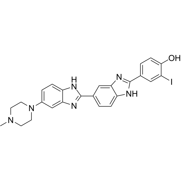
    Hoechst 33342 analog 2

    上海金畔生物科技有限公司为生命科学和医药研发人员提供生物活性分子抑制剂、激动剂、特异性抑制剂、化合物库、重组蛋白,专注于信号通路和疾病研究领域。

    Hoechst 33342 analog 2 

    Hoechst 33342 analog 2 是 Hoechst 33342 的一种类似物, Hoechst 33342 结合到 DNA 小沟,用于细胞 DNA 荧光染色。

    Hoechst 33342 analog 2

    Hoechst 33342 analog 2 Chemical Structure

    CAS No. : 106050-84-4

    规格 价格 是否有货
    10 mM * 1 mL in DMSO ¥4238 询问价格 & 货期
    5 mg ¥3500 询问价格 & 货期
    10 mg ¥5000 询问价格 & 货期
    50 mg ¥15000 询问价格 & 货期

    * Please select Quantity before adding items.

    Hoechst 33342 analog 2 的其他形式现货产品:

    Hoechst 33342 Hoechst 33342 trihydrochloride Hoechst 33342 analog Hoechst 33342 analog 2 trihydrochloride

    生物活性

    Hoechst 33342 analog 2 is an anglog of Hoechst 33342, which is a DNA minor groove binder used fluorochrome for visualizing cellular DNA.

    分子量

    550.39

    Formula

    C25H23IN6O

    CAS 号

    106050-84-4

    运输条件

    Room temperature in continental US; may vary elsewhere.

    储存方式

    4°C, protect from light

    *In solvent : -80°C, 6 months; -20°C, 1 month (protect from light)

    溶解性数据
    In Vitro: 

    DMSO : ≥ 57 mg/mL (103.56 mM)

    * “≥” means soluble, but saturation unknown.

    配制储备液
    浓度 溶剂体积 质量 1 mg 5 mg 10 mg
    1 mM 1.8169 mL 9.0845 mL 18.1689 mL
    5 mM 0.3634 mL 1.8169 mL 3.6338 mL
    10 mM 0.1817 mL 0.9084 mL 1.8169 mL

    *

    请根据产品在不同溶剂中的溶解度选择合适的溶剂配制储备液;一旦配成溶液,请分装保存,避免反复冻融造成的产品失效
    储备液的保存方式和期限:-80°C, 6 months; -20°C, 1 month (protect from light)。-80°C 储存时,请在 6 个月内使用,-20°C 储存时,请在 1 个月内使用。

    参考文献
    • [1]. Chazotte B. Labeling nuclear DNA with hoechst 33342. Cold Spring Harb Protoc. 2011 Jan 1;2011(1):pdb.prot5557.

    所有产品仅用作科学研究或药证申报,我们不为任何个人用途提供产品和服务